
What Is a 9-Week Ultrasound?
A 9-week ultrasound is a pivotal part of early pregnancy care. It falls within the first trimester, typically between 7 to 13 weeks, and is usually recommended to confirm the viability of the pregnancy, assess fetal development, and detect potential concerns early on.
By the 9th week of pregnancy, your embryo is well into the transition to a fetus. Whether performed via a transvaginal probe or abdominal scan, this ultrasound provides a reassuring glimpse into the uterus, offering visible proof of a growing life and a beating heart.
Healthcare providers use the 9-week ultrasound to:
- Confirm intrauterine pregnancy
- Assess fetal heartbeat
- Measure Crown-Rump Length (CRL)
- Evaluate the gestational and yolk sacs
- Check for multiple pregnancies
- Date the pregnancy accurately
At this stage, parents often experience a powerful emotional response; seeing the baby’s shape for the first time brings a sense of reality and joy to the journey ahead.
🧬 Related Topic:
We suggest you read this article.How Many Ultrasounds Are Needed During Pregnancy?
What Can Be Seen in the 9th Week?
With modern devices like the Sono Mobile™ C6, high-resolution imaging makes it possible to detect multiple features clearly at 9 weeks. Thanks to point-of-care ultrasound (POCUS) and portable devices, early pregnancy monitoring is more accessible than ever.
Here’s what a sonographer typically looks for at this point:
Fetal Pole and Crown-Rump Length (CRL)
By week 9, the fetal pole is well-defined and measurable. The CRL is the most reliable parameter for estimating gestational age at this stage. Typical CRL at 9 weeks ranges between 20 to 30 mm.
Fetal Heartbeat
The fetal heart is fully formed and clearly visible. You’ll likely see a rapid flickering movement; a reassuring sign of life. The normal heart rate for a 9-week fetus ranges from 140 to 180 beats per minute.
Gestational Sac and Yolk Sac
The gestational sac should be visible with well-defined borders. Within it, the yolk sac may still be present, although it starts to regress by this time.
Fetal Anatomy Landmarks
At this point, the fetus may begin subtle movements. The head, limb buds, spine, and tiny arms and legs can sometimes be detected, especially with high-resolution ultrasound like Sono Mobile™ C6.
Placental Location
Though still early, placental tissue starts forming and may be visualized along the uterine wall.
Key Features of the Fetus at 9 Weeks

Understanding fetal development at week 9 helps parents and clinicians align expectations and monitor healthy progression. Here are the primary characteristics of the fetus during this week:
Brain and Head
- The head is proportionally large, making up nearly half the fetal length.
- Brain structures are rapidly developing, though still not fully segmented.
Heart
- The four chambers of the heart are formed and functioning.
- You can detect the heartbeat clearly on a good-quality scan.
Limbs and Movement
- Limb buds elongate into arms and legs.
- Tiny toes and fingers begin forming, although webbed.
- First reflexive movements may be detected, though usually not felt by the mother.
Organs
- Major organs, including the liver, kidneys, lungs, and intestines, continue forming.
- The liver begins producing red blood cells.
Size
- Fetal size at week 9 is approximately 2.3 to 3 cm (CRL).
- Weight is around 1 gram — about the size of a grape.
What Happens During the Scan?
Most 9-week ultrasounds are conducted as routine early pregnancy scans, often during the first prenatal appointment. Your OB/GYN or sonographer will determine whether to use a transvaginal or transabdominal approach depending on visibility and patient comfort.
Transvaginal Ultrasound
- Preferred in early pregnancy for better clarity.
- A small probe is gently inserted into the vaginal canal.
- Provides clearer images of the uterus and embryo.
Transabdominal Ultrasound
- May be used if the uterus is clearly visible through the abdomen.
- Requires a full bladder to help visualize the uterus.
- Less invasive and generally more comfortable.
Steps During the Scan:
- Patient Preparation; You may be asked to drink water and not urinate.
- Image Acquisition; The sonographer will position the probe and adjust settings.
- Measurement; Key dimensions like CRL and gestational sac diameter will be measured.
- Doppler Evaluation (optional); May be used to detect fetal heartbeat.
- Counseling; The doctor reviews results and answers any questions.
🧬 Related Topic:
Determining the sex of the fetus is done in various ways, and you can read about the best method, Finding Out Baby’s Sex With an Ultrasound, in this article.
Common Questions About the 9-Week Scan
Is a heartbeat always visible at 9 weeks?
In most cases, yes. The fetal heart should be beating clearly, and absence of a heartbeat may prompt further evaluation. If your cycles are irregular, the pregnancy may be younger than expected.
What if the measurements are smaller than expected?
A small CRL could indicate incorrect dating, especially if ovulation occurred later. Your provider may schedule a follow-up scan.
Can abnormalities be detected at this stage?
Major structural anomalies are difficult to identify at week 9. However, missing heartbeat, abnormal yolk sac, or irregular gestational sac shape can raise concerns and require monitoring.
Will I get a picture?
Yes, most clinics offer a printed image or digital file of the scan. Using modern handheld devices like Sono Mobile™ C6, clinicians can even capture high-resolution snapshots on the spot.
Is the scan safe for the baby?
Absolutely. Ultrasound uses sound waves, not radiation, and is widely accepted as safe during all stages of pregnancy.
When is the next scan after 9 weeks?
Usually at 12 weeks, when the first-trimester screening for chromosomal anomalies (nuchal translucency) is done.
The Role of Sono Mobile™ in Early Pregnancy Scans
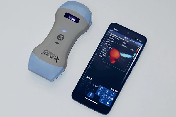
At Fagonex, we understand the power of bringing imaging closer to the point of care. The Sono Mobile™ handheld ultrasound system is engineered to meet the needs of both clinicians and patients; especially in rural, emergency, and home-care settings.
With the Sono Mobile™ C6, obstetricians and midwives can:
- Conduct 9-week ultrasounds on the go
- Offer care in underserved areas without bulky equipment
- Immediately detect heartbeat and fetal position
- Capture and store high-resolution fetal images
- Provide fast diagnostics in high-volume prenatal clinics
Who Benefits from Point-of-Care Ultrasound?
- Expecting mothers seeking reassurance in early pregnancy
- Midwives working in remote or mobile setups
- Primary care providers performing routine prenatal visits
- Healthcare teams in ERs and urgent care centers
With Sono Mobile™ C6, early scans such as the 9-week ultrasound become more accessible, affordable, and time-efficient; no waiting for appointments, no delays in reassurance.
Conclusion: What a 9-Week Ultrasound Means
A 9-week ultrasound is much more than a routine check; it’s a window into the earliest stage of life, offering reassurance, insight, and connection. For patients, it’s often the first time they see their baby’s heartbeat. For providers, it’s an essential tool in confirming and monitoring healthy fetal development.
Using cutting-edge tools like Sono Mobile™ C6 from Fagonex, clinicians can provide real-time diagnostics, reduce patient anxiety, and enhance maternal-fetal care; all while remaining portable, accurate, and professional.
Whether you’re a new parent, a healthcare student, or a provider seeking better imaging tools, understanding the 9-week ultrasound empowers you to appreciate one of the most critical milestones in the journey of pregnancy.
📍 Explore more about point-of-care obstetric imaging and our handheld solutions at https://fagonex.com/product-category/handheld-ultrasound/
Contact us to learn how Sono Mobile™ can support your clinical practice.
One Response
I really appreciate this post. I¦ve been looking everywhere for this! Thank goodness I found it on Bing. You’ve made my day! Thx again