
Point-of-Care Ultrasound in Anesthesia
Point-of-care ultrasound (POCUS) can support anesthesiologists with real-time visualization during selected procedures and focused perioperative assessments. From regional anesthesia and vascular access to lung, cardiac, airway, gastric and other focused applications, ultrasound can bring relevant anatomical information directly to the bedside, operating room, procedure area or recovery environment.
When performed by appropriately trained clinicians, anesthesia ultrasound can complement physical examination and other clinical information while supporting procedural planning and focused clinical questions. For a broader introduction, explore our Point-of-Care Ultrasound (POCUS) guide .
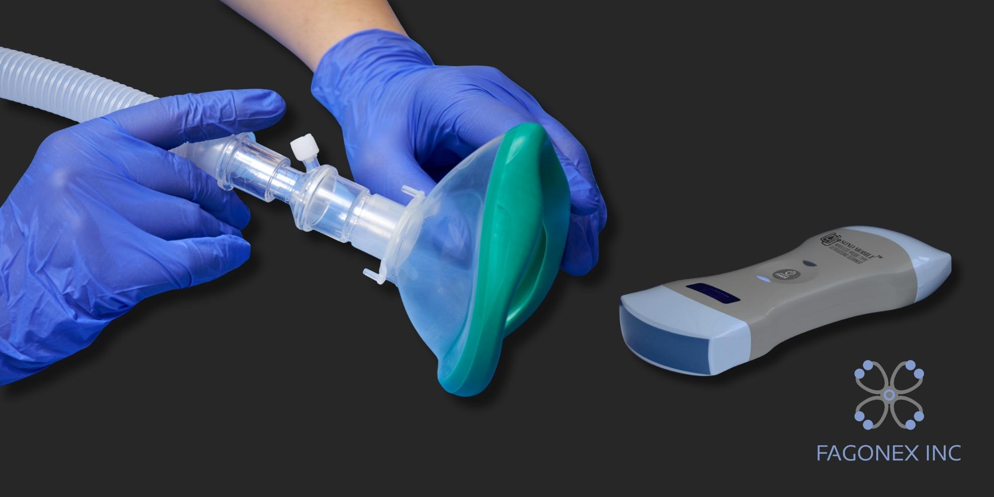
Regional Anesthesia and Ultrasound-Guided Nerve Blocks
Ultrasound-guided regional anesthesia allows the clinician to visualize relevant anatomy before and during selected procedures. Depending on the clinical application, ultrasound may help identify peripheral nerves, fascial planes, muscles, blood vessels and surrounding soft tissues.
Real-Time Sonoanatomy
Ultrasound provides dynamic visualization of anatomical structures, helping clinicians understand the relationship between the intended target and nearby vessels, muscles and soft tissues.
Needle Guidance
During appropriately selected image-guided procedures, ultrasound may support visualization of the planned needle path and surrounding anatomy.
- Peripheral nerve blocks
- Selected fascial plane blocks
- Identification of target anatomy
- Needle visualization during selected procedures
- Assessment of anatomical variation
Ultrasound-Guided Vascular Access
Ultrasound-guided vascular access is an important procedural application in anesthesiology, perioperative medicine and critical care . Real-time imaging can help clinicians visualize vessel location, depth, surrounding structures and needle position during selected vascular-access procedures.
Potential Applications
- Peripheral vascular access
- Central venous access
- Arterial line placement
- Difficult vascular access
- Vessel and surrounding-anatomy visualization
Why Real-Time Imaging Matters
Direct visualization may support procedural planning when surface landmarks are limited or anatomy is difficult to assess clinically. Appropriate sterile technique, training and institutional protocols remain essential.
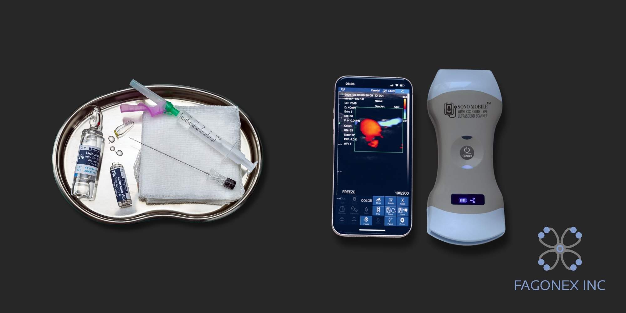
Perioperative Point-of-Care Ultrasound
Beyond procedural guidance, point-of-care ultrasound in anesthesia can help clinicians address selected perioperative clinical questions at the bedside. The examination should remain focused, interpreted in clinical context and performed within the operator's training and institutional scope.
Focused Cardiac Ultrasound
Selected cardiac views may support focused assessment of global cardiac activity, gross ventricular appearance, pericardial fluid and other perioperative questions when clinically appropriate. In patients with undifferentiated hypotension or shock, clinicians may also use the RUSH exam when appropriate.
Lung & Pleural Ultrasound
Lung ultrasound may support assessment of lung sliding, pleural fluid, selected interstitial patterns and other focused respiratory findings.
Airway Ultrasound
Ultrasound may support selected evaluation of airway anatomy and procedural planning in appropriately trained hands.
Gastric & Preoperative Assessment
In selected circumstances, focused gastric and other preoperative ultrasound examinations may contribute additional information to the broader perioperative assessment.
The American Society of Regional Anesthesia and Pain Medicine (ASRA) maintains a dedicated perioperative POCUS community focused on advancing education and knowledge in this field. Explore ASRA perioperative POCUS resources .
Anesthesia POCUS Video Examples
Explore real-time ultrasound examples relevant to anesthesiology, perioperative point-of-care ultrasound, vascular assessment, airway evaluation and focused cardiac ultrasound . These examples demonstrate how bedside ultrasound can provide immediate anatomical and physiological information across different clinical applications.
Femoral Vessels Ultrasound
Real-time ultrasound visualization of the femoral vascular anatomy, relevant to vascular assessment and ultrasound-guided access in anesthesia and perioperative care.
Inferior Vena Cava (IVC) Ultrasound
Focused visualization of the inferior vena cava as part of selected hemodynamic and volume-related POCUS assessments when clinically appropriate.
Parasternal Long Axis (PLAX) View
A focused cardiac ultrasound example demonstrating the parasternal long-axis view, one of the commonly used windows in bedside cardiac POCUS.
Vocal Cord Ultrasound & Airway Assessment
Ultrasound visualization of vocal-cord movement as an example of selected airway ultrasound applications relevant to perioperative and anesthesia assessment.
Why Handheld Ultrasound Fits Modern Anesthesia Workflows
Handheld ultrasound can bring focused imaging directly to the clinical environment without relying on a fixed cart-based system. This may be useful across operating rooms, preoperative areas, post-anesthesia care units, critical care units, pain clinics and procedure rooms.
Portable Access
Compact wireless systems can support bedside deployment when clinicians need rapid access to focused imaging or procedural guidance.
Non-Ionizing Imaging
Ultrasound uses sound waves rather than ionizing radiation and may be repeated when clinically appropriate.
Education & Training
Portable systems may support hands-on education in sonoanatomy, vascular access, regional anesthesia, lung ultrasound and focused cardiac ultrasound.
Challenging Anatomy
Real-time visualization may provide additional anatomical information when conventional landmarks are difficult to identify.

Sono Mobile® CL64 for Anesthesiology
Sono Mobile® CL64 is a wireless dual-head handheld ultrasound system designed to support a broad range of point-of-care ultrasound applications in anesthesiology. Its convex and linear transducers, combined with a phased-array preset, allow clinicians to use one portable platform across selected procedural, cardiac, lung, vascular and general bedside ultrasound applications.
Probe Configuration
- Dual-head convex and linear transducers
- Phased-array preset for selected cardiac applications
- Frequency range from 3.2 to 10 MHz
- Designed for superficial and deeper imaging applications
Imaging & Workflow
- B, B/M, Color, PDI and PW imaging modes
- Wireless handheld workflow
- Compatible with supported mobile, tablet and Windows devices
- No recurring application subscription fee
For anesthesiology teams, CL64 can support selected applications such as regional anesthesia, vascular access, lung ultrasound, focused cardiac ultrasound and bedside procedural guidance, depending on clinician training, institutional protocols and the clinical indication.
Using Ultrasound Responsibly in Anesthesia Practice
Point-of-care ultrasound should be used by appropriately trained healthcare professionals and interpreted together with the patient's history, physical examination and other clinically indicated diagnostic information. A focused bedside ultrasound examination does not replace comprehensive diagnostic imaging, specialist interpretation or additional testing when these are required.
Clinical applications, credentialing requirements, documentation standards and infection-control practices may vary by institution and jurisdiction. Users should follow applicable professional guidance, local policies and competency requirements.
Anesthesia Ultrasound FAQ
How is ultrasound used in anesthesiology?
Appropriately trained anesthesiologists may use ultrasound for selected applications including regional anesthesia, nerve blocks, vascular access, focused cardiac and lung assessment, airway evaluation and other perioperative POCUS examinations.
Can handheld ultrasound be used for regional anesthesia?
Handheld ultrasound may support visualization of nerves, fascial planes, vessels and surrounding anatomy during selected ultrasound-guided regional anesthesia procedures when the device, probe configuration and operator training are appropriate for the intended application.
Which Sono Mobile® model is suitable for anesthesia?
Sono Mobile® CL64 is designed for broad POCUS use and combines convex and linear transducers with a phased-array preset. This configuration may support selected regional anesthesia, vascular, lung, cardiac and general bedside ultrasound applications.
Does POCUS replace formal diagnostic ultrasound?
No. POCUS is a focused examination intended to answer specific clinical questions at the point of care. Formal diagnostic imaging or specialist interpretation may still be required depending on the clinical situation.
Bring Point-of-Care Ultrasound Into Your Anesthesia Workflow
Explore Sono Mobile® CL64 for regional anesthesia, vascular access, perioperative POCUS, bedside assessment and ultrasound education.