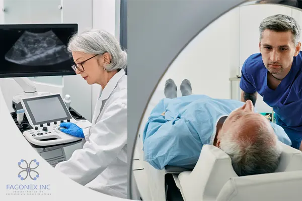
In the world of diagnostic imaging, ultrasound and CT (computed tomography) scans are two of the most commonly used tools. Each has its unique strengths and uses, helping medical professionals to diagnose and treat a wide range of health conditions. By understanding the differences between ultrasound and CT scans, patients and healthcare providers alike can better choose the best imaging technique for each situation.
Whether it’s examining a developing fetus, diagnosing a tumor, or assessing a traumatic injury, both ultrasound and CT scans play essential roles. This article will explore these two imaging methods, breaking down their distinct features, uses, and how each fits into modern medical practices.
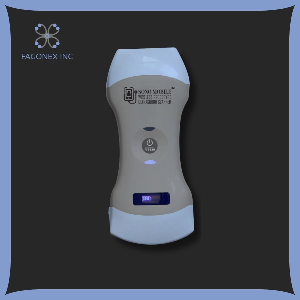
What is Handheld Ultrasound?

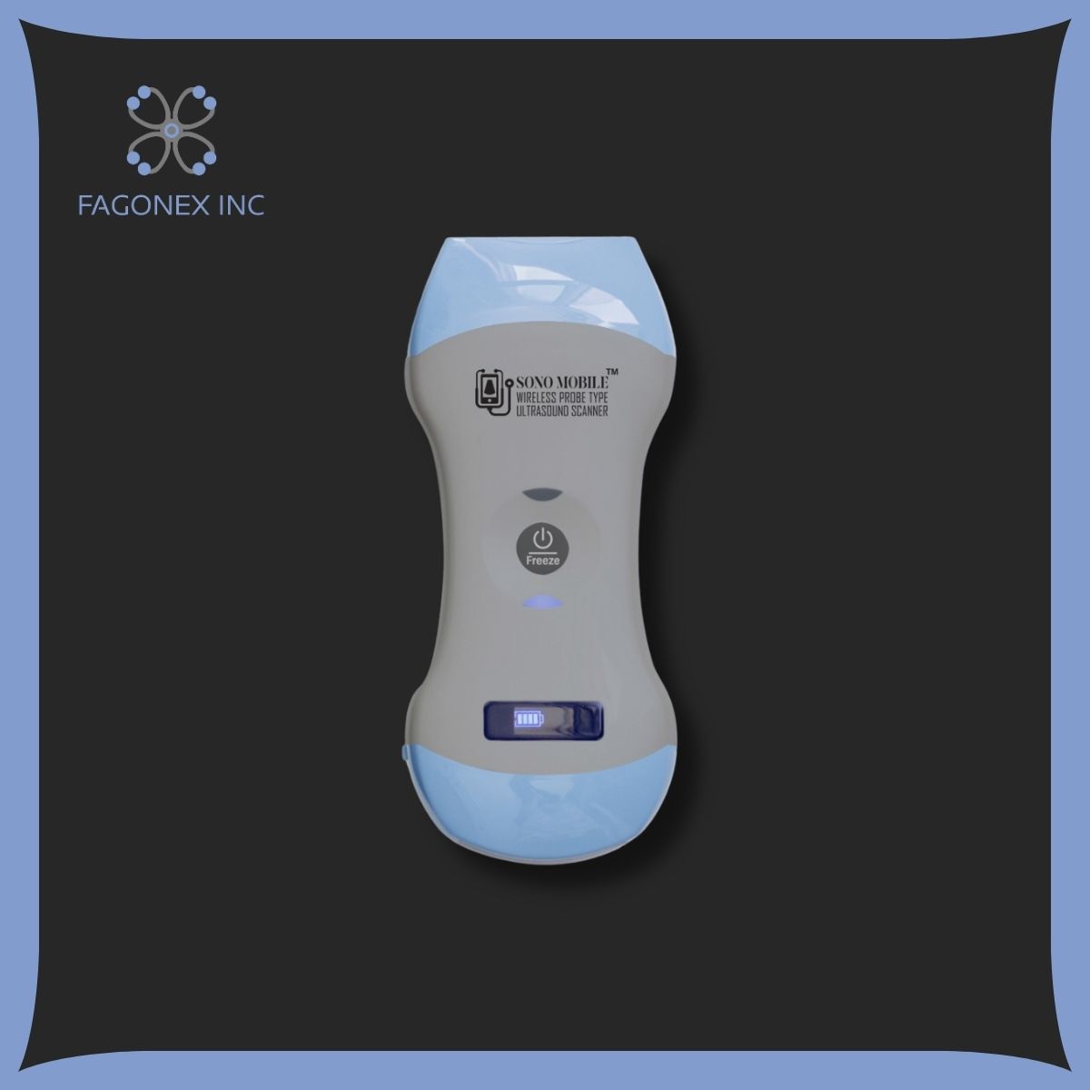
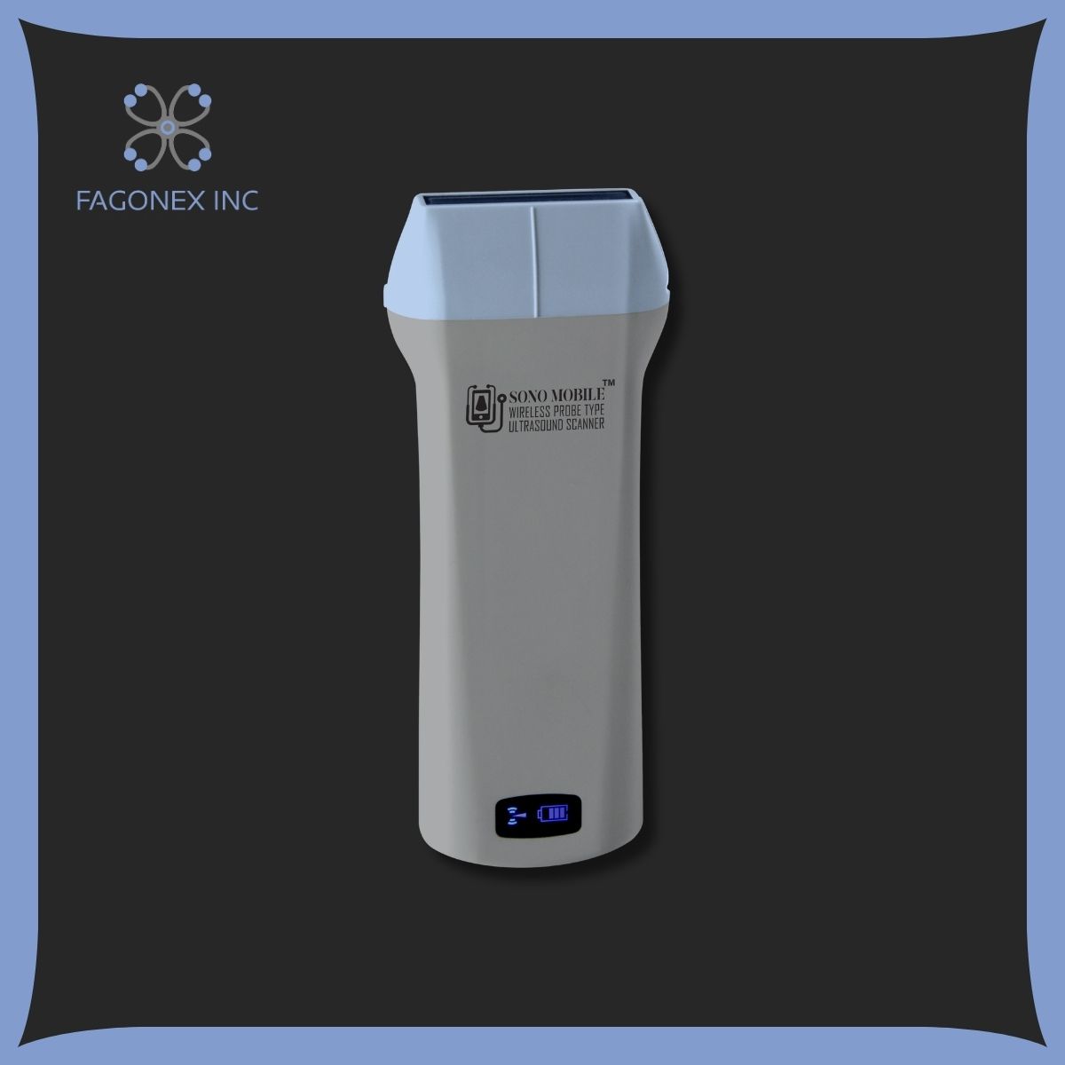
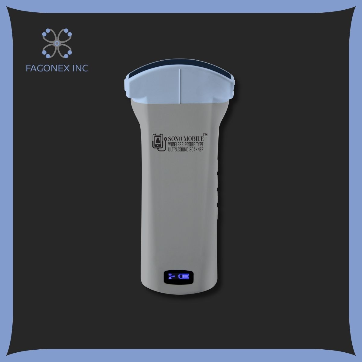
Handheld ultrasound devices are compact, portable machines that offer many of the capabilities of traditional ultrasound equipment with the convenience of mobility. Unlike large ultrasound machines commonly found in hospitals, handheld devices allow healthcare providers to deliver imaging directly to the patient, whether in a hospital, clinic, or remote setting.
Handheld ultrasound works by sending high-frequency sound waves into the body, which then bounce off structures and return as echoes. These echoes are captured and translated into real-time images, allowing practitioners to monitor body structures and functions instantly.
Read More: Reasons to choose Handheld Ultrasound
Some common applications of handheld ultrasound include:
- Emergency Situations: In trauma cases, responders use handheld ultrasound to quickly assess conditions like internal bleeding or organ injury. Having a portable ultrasound can make a crucial difference in remote or rural areas where larger machines aren’t accessible.
- Bedside Monitoring: Doctors and nurses often use handheld ultrasound devices for bedside evaluations, especially for conditions requiring continuous monitoring like congestive heart failure.
- Field and Remote Medicine: Portable ultrasound devices are useful in rural areas and during medical missions, where access to traditional imaging machines is limited.
Handheld ultrasounds are also revolutionizing healthcare by making diagnostic imaging more accessible and affordable. As technology improves, handheld devices are closing the gap with traditional ultrasound machines, becoming an essential tool in modern medicine.
Read More: What Are Portable Ultrasound Machines+types and Benefits
What Is a CT Scan?

A CT scan, or computed tomography scan, is a type of imaging that uses X-rays and a computer to create cross-sectional images of the body. Unlike traditional X-rays that capture only a single view, CT scans take multiple images from different angles, which are then combined to produce detailed, three-dimensional pictures of the body’s internal structures.
CT scans provide higher-resolution images than ultrasounds, making them particularly useful for diagnosing complex conditions. For example, a CT scan of the head might help detect subtle fractures or bleeding that could be life-threatening, while a scan of the chest can reveal detailed views of the lungs and heart.
CT scans are especially effective for:
- Detecting Tumors and Cancer: By providing a detailed view of tissues, CT scans are essential for locating and sizing tumors, as well as monitoring them over time.
- Guiding Surgeries: Surgeons use CT scans to plan procedures, helping to map out areas like tumors or blood vessels in detail.
- Assessing Internal Injuries: After a traumatic accident, CT scans are often the first choice for assessing internal injuries, as they offer a comprehensive view of the body’s organs, bones, and blood vessels.
The process of a CT scan involves the patient lying on a motorized table that slides into a cylindrical machine. The scanner rotates around the patient, capturing images that the computer then combines into a detailed, 3D image. Though CT scans involve exposure to low levels of radiation, they are considered safe and invaluable for situations that require detailed imaging.
Difference Between Ultrasound and CT Scan
When deciding between an ultrasound and a CT scan, understanding the unique characteristics of each can help make the most informed choice. Here’s a closer look at their primary differences:
Technology and Imaging Type
- Ultrasound: Uses sound waves to create images of organs, tissues, and blood flow. Ultrasound is ideal for soft-tissue imaging, such as examining a fetus during pregnancy or viewing a patient’s heart in real-time. It’s also a safer option for procedures requiring repeated imaging.
Read More: Facts about Ultrasound
- CT Scan: Uses X-rays to capture multiple angles and combines these images to create cross-sectional, high-resolution views of internal body structures. CT scans are best for detailed imaging of bones, organs, and other complex structures, making them particularly valuable for assessing traumatic injuries and planning surgeries.
Examples of Use
- Ultrasound Example: A physician may use ultrasound to view the development of a fetus or to check for blood flow issues in the veins and arteries.
- CT Scan Example: CT scans are the preferred choice for detecting abnormalities in the brain, such as strokes or tumors, where clear, detailed images are critical.
Level of Detail and Accuracy
Ultrasounds and CT scans differ in the clarity and type of information they provide.
- Ultrasound Detail: Ultrasound is effective for visualizing real-time motion, which makes it ideal for observing a heartbeat, fetal movement, or blood flow. However, it’s generally less detailed than a CT scan and may struggle with imaging deeper body structures, especially if air or gas is present.
- CT Scan Detail: CT scans offer a higher level of detail, especially useful for viewing internal organs and complex structures like the brain and lungs. This level of accuracy is critical for diagnosing conditions that require in-depth anatomical views, such as cancers or serious internal injuries.
Safety and Radiation Exposure
- Safety of Ultrasound: Ultrasound is considered one of the safest imaging techniques as it uses sound waves rather than radiation, making it suitable for pregnant women and routine check-ups.
- Radiation in CT Scans: CT scans do involve a small amount of radiation exposure. While this is not typically harmful in limited use, the risks may increase with frequent scans, so physicians generally use CT only when necessary.
Speed and Accessibility
- Handheld Ultrasound Accessibility: Ultrasound exams are usually quicker and can be conducted in a wide range of settings, including clinics and even at the patient’s bedside. Handheld ultrasound devices, like those offered by Fagonex, enhance accessibility by allowing healthcare professionals to provide imaging wherever it’s needed, often within minutes.
- CT Scan in Hospitals: CT scans are typically performed in hospitals or specialized imaging centers, as the equipment is large and complex. The entire procedure can take longer than an ultrasound, although advanced CT machines are faster than ever.
Cost and Affordability
- Ultrasound: Ultrasounds tend to be more affordable than CT scans, as they don’t require complex equipment and use sound waves instead of X-ray technology.
- CT Scan: CT scans are generally more expensive due to the sophisticated equipment and the need for specially trained technicians.
Uses of Handheld Ultrasound

Handheld ultrasounds are extremely versatile and find use in various medical applications. Their portability and accessibility make them invaluable tools in:
- Emergency and Trauma Care: Handheld ultrasounds are used to assess injuries quickly, especially in remote or emergency settings.
- Cardiology: Doctors use handheld ultrasounds to examine heart function, monitor blood flow, and assess cardiovascular health.
- OB-GYN: Obstetricians use handheld ultrasound devices to monitor fetal development throughout pregnancy, including tracking growth and heartbeat.
To learn more about buying a handheld ultrasound, and best portable ultrasound devices from Fagonex, visit our product page.
Uses of CT Scan
CT scans are indispensable for conditions that require in-depth and high-resolution imaging. Here are some of their primary uses:
- Oncology: CT scans help detect, measure, and monitor tumors, making them essential for cancer diagnosis and treatment planning.
- Neurology: Brain CT scans are used to detect issues like strokes, brain tumors, and traumatic injuries.
- Orthopedics: CT scans provide clear images of bones and joints, which help diagnose fractures and other orthopedic conditions.
Conclusion
Both ultrasound and CT scans have unique strengths and applications. Handheld ultrasounds offer a safe, accessible, and portable imaging solution, ideal for emergency situations, bedside monitoring, and various specialties. CT scans, on the other hand, provide higher-resolution images that are invaluable for diagnosing complex internal conditions and planning surgical interventions.
When deciding between these two methods, it’s essential to consider factors such as the type of condition being assessed, the level of detail required, safety, and accessibility. With technology advancing rapidly, both ultrasound and CT scans are likely to become even more powerful and accessible, continuing to improve patient care around the world.
One Response
Really appreciate this helpful writing.