When it comes to medical imaging, few tools are as commonly used or widely recognized as ultrasound and X-ray. These technologies have revolutionized modern healthcare by allowing doctors to see inside the body without surgery, providing essential insights that guide diagnosis and treatment. Yet, despite their widespread use, ultrasound and X-ray differ significantly in terms of technology, application, and advantages.
Understanding the strengths and limitations of these imaging methods can help patients make informed decisions about their care. This article will delve into the fundamentals of handheld ultrasound and X-ray imaging, comparing their uses, benefits, and risks. By the end, you’ll have a clearer understanding of which method might be most appropriate for different medical needs, and why both remain indispensable in today’s medical field.
What Is Handheld Ultrasound?

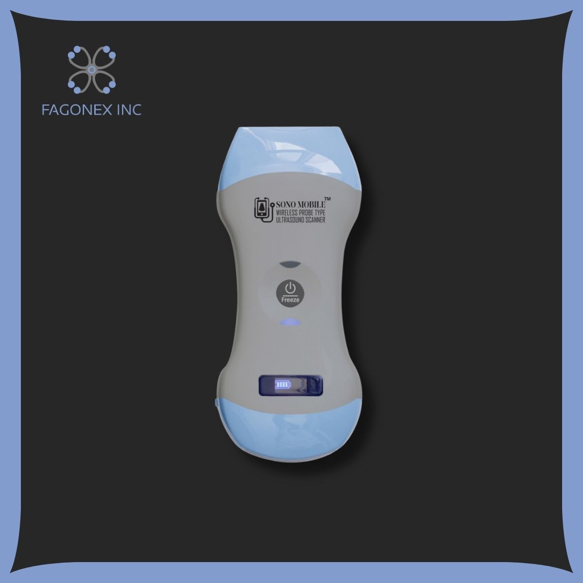
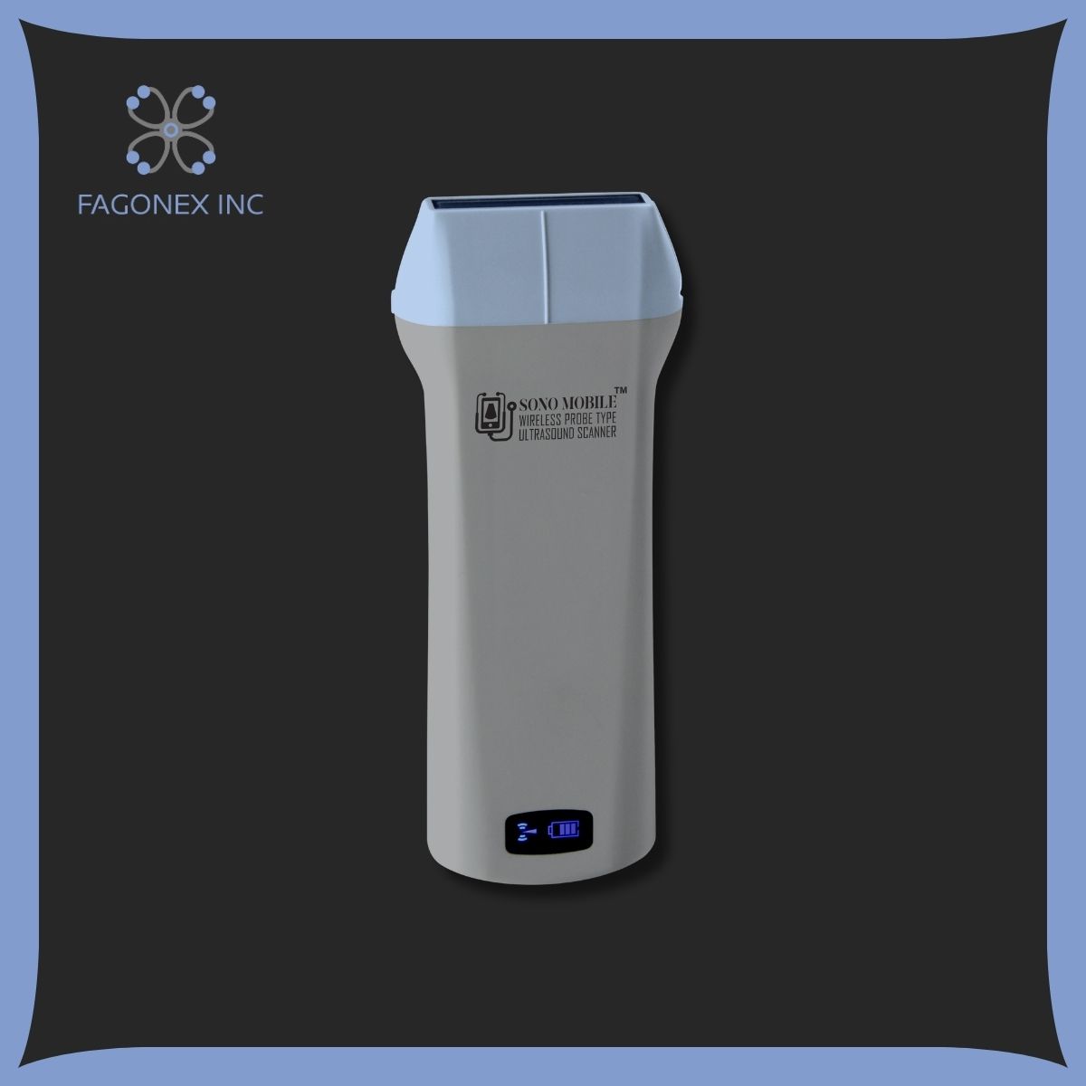
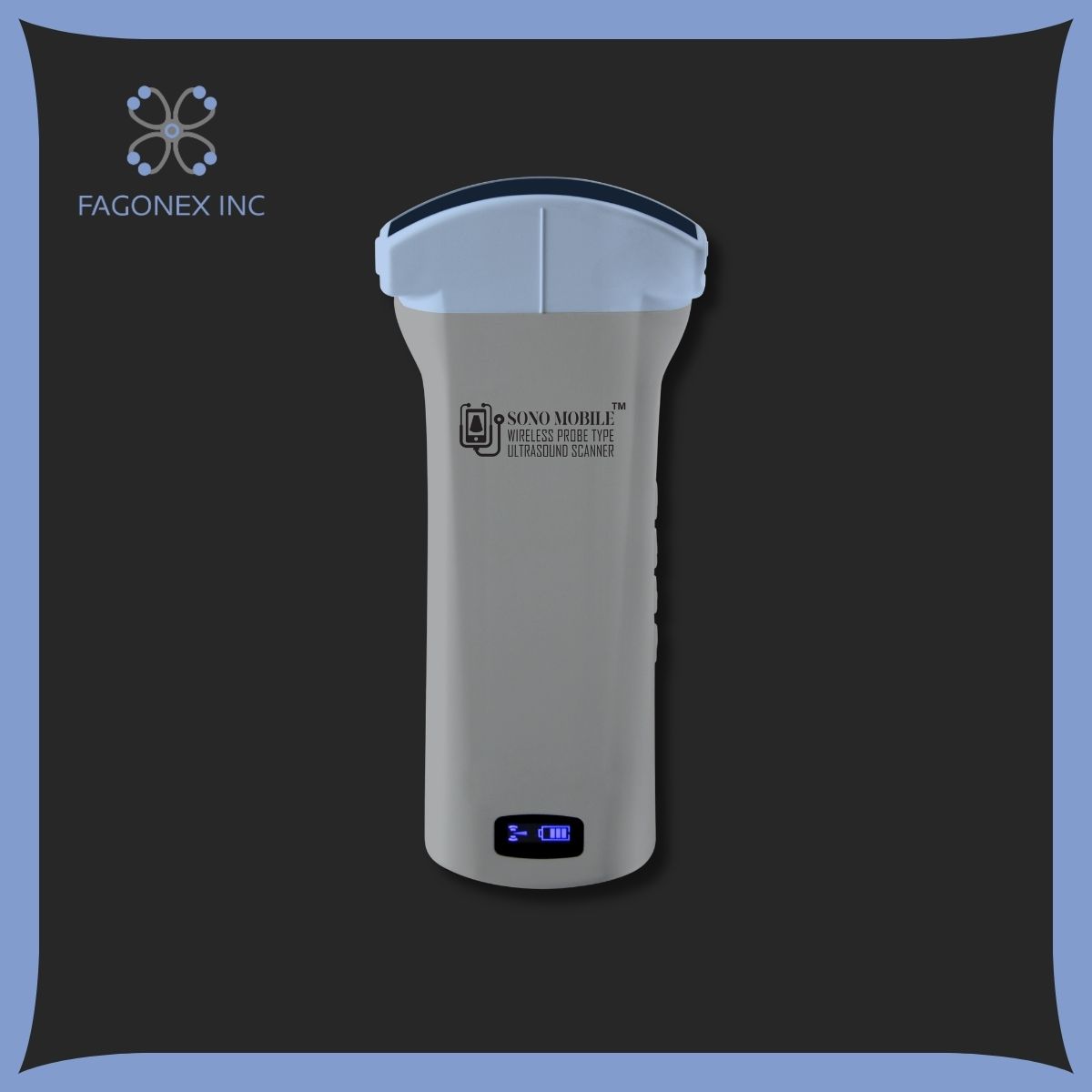
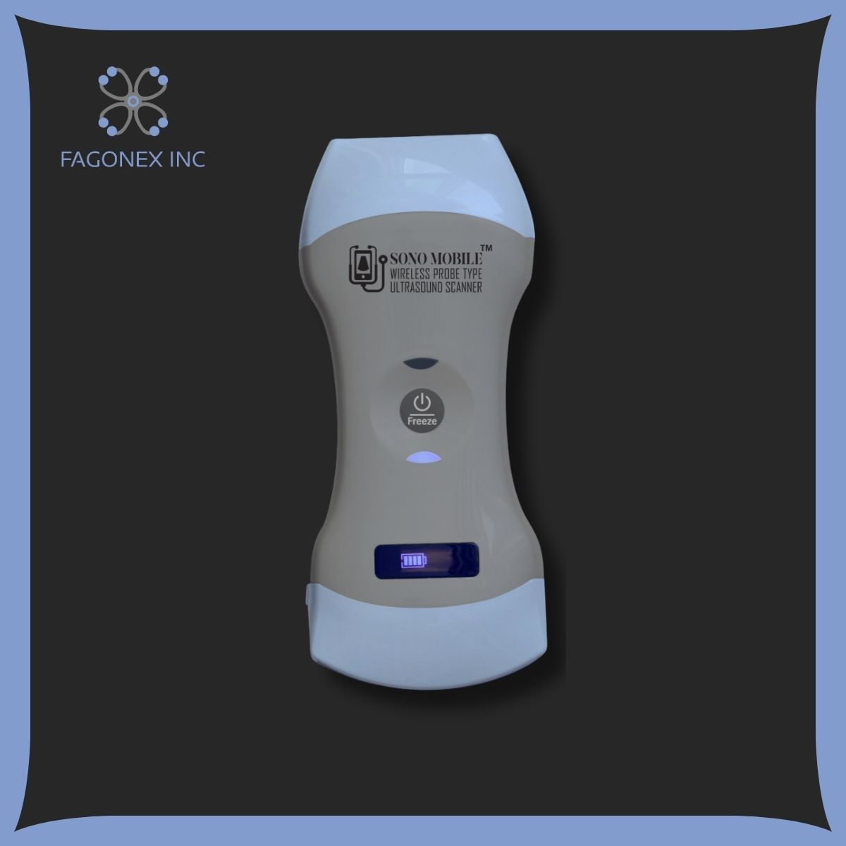
Handheld ultrasound, also known as portable ultrasound, represents a significant advance in medical imaging. Unlike traditional ultrasound machines, which are large and often confined to hospitals, handheld ultrasound devices are small, mobile, and can be used in various settings. They’re designed for portability, making it easier for healthcare providers to bring imaging technology to the bedside or even to remote areas.
Handheld ultrasounds work by emitting high-frequency sound waves that bounce off tissues and organs. These waves create real-time images, allowing doctors to observe internal structures in great detail. One of the biggest benefits of handheld ultrasound is its flexibility; it can be used in emergency departments, intensive care units, outpatient clinics, and more. For example, they’re frequently used in prenatal care, where ultrasounds help to monitor fetal growth and even determine the baby’s sex.
Read More: Finding Out Baby’s Sex With an Ultrasound
Advantages of Handheld Ultrasound
- Portability: Small, lightweight, and battery-powered, these devices are ideal for home healthcare and remote locations.
- Non-Invasive: With no radiation, handheld ultrasound can be used safely in various scenarios, including with pregnant women and children.
- Real-Time Imaging: Portable ultrasound machines offer instant results, making them useful in emergencies and for quick diagnostics.
For those looking to buy handheld ultrasound devices, many options are now available with advanced imaging capabilities, suitable for general practitioners, specialists, and emergency responders alike.
Read More: Best portable Ultrasound Devices
What Is X-Ray?

X-ray imaging, one of the oldest and most trusted imaging methods, has been a cornerstone of medical diagnostics since its discovery in the late 19th century. X-rays use a type of electromagnetic radiation that can pass through the body. The denser the tissue (like bones), the more it absorbs the X-ray, resulting in a white or light gray image on the film. Softer tissues, like muscles and organs, absorb less radiation and thus appear darker, allowing radiologists to differentiate structures within the body.
X-rays are particularly useful for diagnosing bone fractures, assessing chest conditions, and even detecting certain types of cancers. While the radiation dose is generally low, precautions are taken to minimize exposure, especially for children and pregnant women. Lead aprons, for instance, are often used to protect patients from unnecessary radiation.
Types of X-Ray Procedures
- Standard X-Ray: Used for examining bones, joints, and certain soft tissue structures.
- CT Scan (Computed Tomography): An advanced X-ray that provides detailed cross-sectional images, commonly used for complex internal assessments.
Read More: Difference Between Ultrasound and CT Scan
- Mammography: A specialized X-ray procedure to detect early signs of breast cancer.
- Dental X-Ray: Used by dentists to examine teeth, roots, and jawbones.
Each of these procedures plays a unique role in medical imaging, helping doctors visualize conditions that would otherwise require surgery to diagnose.
Difference Between Ultrasound and X-Ray
While ultrasound and X-ray are both crucial in medical imaging, they operate on entirely different principles and have distinct applications.
- Technology: Ultrasound uses sound waves to create images, which is ideal for soft tissues. In contrast, X-rays rely on electromagnetic radiation to capture images, which excels at visualizing dense structures like bones.
- Safety: Ultrasound is generally considered safer because it doesn’t involve radiation. This makes it the preferred choice for frequent imaging, especially in pregnancy and pediatrics. X-rays, however, use ionizing radiation, and while safe in limited doses, are less suitable for repetitive use.
- Applications: Ultrasound is used for soft tissue examination, such as the heart, kidneys, liver, and muscles. X-rays are essential for hard structures, like bones and certain organs, and are particularly useful in assessing injuries and fractures.
- Portability: Handheld ultrasounds offer unprecedented mobility, allowing for real-time imaging in virtually any location. X-ray machines are generally less portable, though mobile X-ray units do exist for hospital settings.
In summary, each technology serves a unique purpose, and both are essential in the broader landscape of medical diagnostics.
Uses of Handheld Ultrasound

Handheld ultrasound is transforming how healthcare is delivered, especially in areas where traditional, stationary equipment is impractical. Below are some of the key uses for handheld ultrasound devices, which provide versatility for many fields and medical situations:
- Emergency Situations: In trauma settings, handheld ultrasound can quickly detect internal bleeding, fluid buildup, or organ damage.
- Point-of-Care Testing (POCT): Handheld ultrasound allows doctors to perform immediate imaging in various clinical settings, from the bedside to outpatient clinics.
- Guided Procedures: Ultrasounds are often used to guide procedures like needle aspirations, biopsies, and injections with precision.
- Cardiology: Portable ultrasound machines allow cardiologists to assess heart function, measure blood flow, and identify issues like valve abnormalities.
- Abdominal Scans: Ultrasounds can be used to evaluate abdominal organs, identify stones in the gallbladder or kidneys, and assess other internal conditions.
For healthcare providers or organizations seeking more affordable and flexible imaging solutions, investing in portable ultrasound machines like those offered by Fagonex can make a significant difference.
Read More: What Are Portable Ultrasound Machines+types and Benefits
What Can Ultrasound Diagnose?

Ultrasound is highly effective in diagnosing a variety of conditions, particularly those involving soft tissues or organs. Here are several examples:
- Pregnancy: Ultrasound is invaluable in monitoring fetal development, assessing amniotic fluid levels, and determining the baby’s position. It is also the primary method for finding out the baby’s sex with an ultrasound, offering a safe, non-invasive option.
- Gallstones and Kidney Stones: These solid formations can be visualized using ultrasound, aiding in early diagnosis and treatment.
- Heart and Blood Vessel Conditions: Echocardiography, a type of ultrasound, is used to evaluate heart health, detecting conditions like heart failure, valve disorders, and abnormal blood flow.
- Thyroid Abnormalities: Ultrasound helps in diagnosing thyroid nodules, enlargement, or cysts.
- Soft Tissue Injuries: Muscles, ligaments, and tendons can be examined using ultrasound to identify tears, inflammation, or other injuries.
As handheld ultrasound technology advances, its applications continue to expand, providing immediate insights that can help guide timely treatment.
What Can X-Ray Diagnose?
X-ray imaging excels in diagnosing conditions that involve bones, teeth, and certain organs. Its ability to capture detailed images of dense structures has made it invaluable in several medical fields:
- Bone Fractures: X-rays are the primary tool for diagnosing fractures, dislocations, and joint damage.
- Spinal Conditions: Spine abnormalities, such as herniated discs or vertebral fractures, are often identified with X-rays.
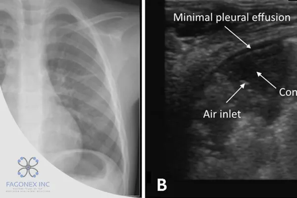
- Lung and Chest Diseases: Chest X-rays are essential in diagnosing pneumonia, tuberculosis, lung tumors, and other respiratory conditions.
- Dental Problems: Dentists rely on X-rays to detect cavities, infections, and other dental issues that aren’t visible during a regular examination.
- Arthritis: X-rays can show joint damage caused by arthritis, helping doctors monitor disease progression and adjust treatment plans.
By providing clear images of bones and hard tissues, X-rays are indispensable for evaluating injuries, infections, and chronic conditions.
Uses of X-Ray
Beyond diagnosing specific conditions, X-ray technology has diverse uses across medical and dental fields:
- Orthopedic Assessments: X-rays are essential for assessing fractures, bone growth, and joint conditions, making them crucial in orthopedic care.
- Dental Imaging: X-rays are frequently used to detect cavities, monitor root health, and guide procedures like root canals.
- Chest Examinations: X-ray imaging is the standard method for diagnosing chest infections, lung diseases, and heart conditions.
- Cancer Screening: Mammography, a type of X-ray, is critical for early breast cancer detection, improving patient outcomes.
- Infection Detection: X-rays can help detect infections like pneumonia, which may not be immediately evident through other methods.
For those interested in medical devices, Fagonex offers advanced ultrasound technology as a complementary tool to X-ray, each playing a unique role in comprehensive patient care.
Conclusion
Ultrasound and X-ray imaging are two pillars of modern diagnostics, each with distinct strengths. Ultrasound provides a safe, radiation-free method for imaging soft tissues and is often used for real-time assessments of organs and blood flow. X-rays, on the other hand, excel at imaging bones and dense structures, making them essential for diagnosing fractures, joint issues, and chest conditions.
As medical technology advances, tools like handheld ultrasound are becoming more accessible, allowing for quick, convenient imaging in nearly any setting. The synergy between ultrasound and X-ray imaging enables healthcare providers to offer faster, more accurate diagnoses, ultimately improving patient care.