What Is a Pelvic Ultrasound?
A pelvic ultrasound is a non-invasive diagnostic imaging technique that uses high-frequency sound waves to generate real-time images of the organs and structures within the lower abdomen and pelvic cavity. It is commonly used in gynecology, urology, and internal medicine to examine the uterus, ovaries, fallopian tubes, bladder, prostate, and surrounding tissues.
Unlike CT scans or X-rays, pelvic ultrasound does not involve radiation, making it a safe option for repeated use and during pregnancy. Whether to evaluate pelvic pain, irregular menstrual bleeding, urinary retention, infertility, or early pregnancy complications, this procedure provides accurate, timely, and painless insight into internal conditions.
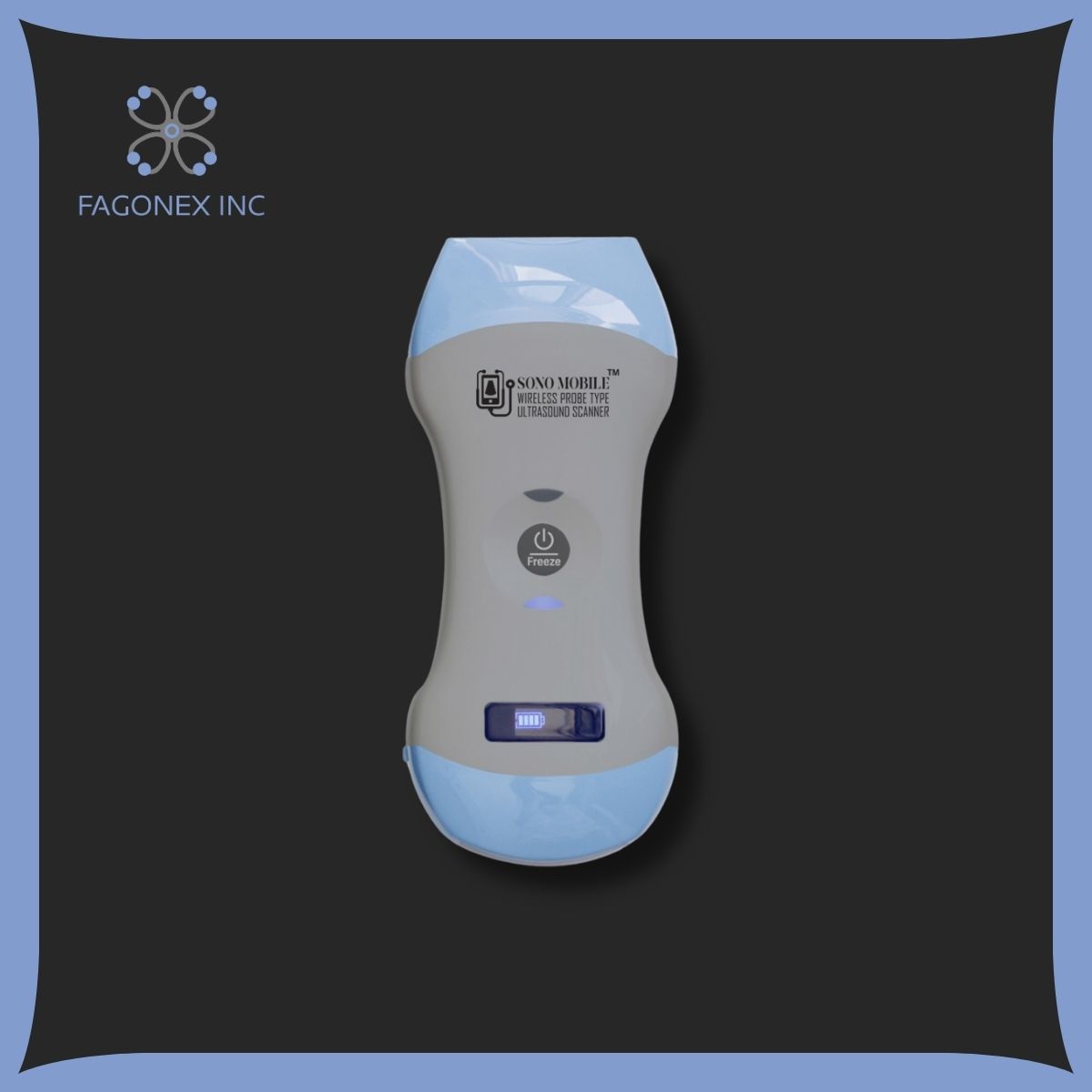
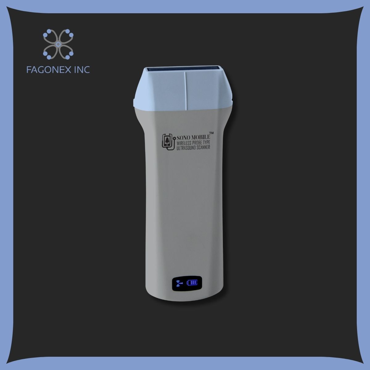
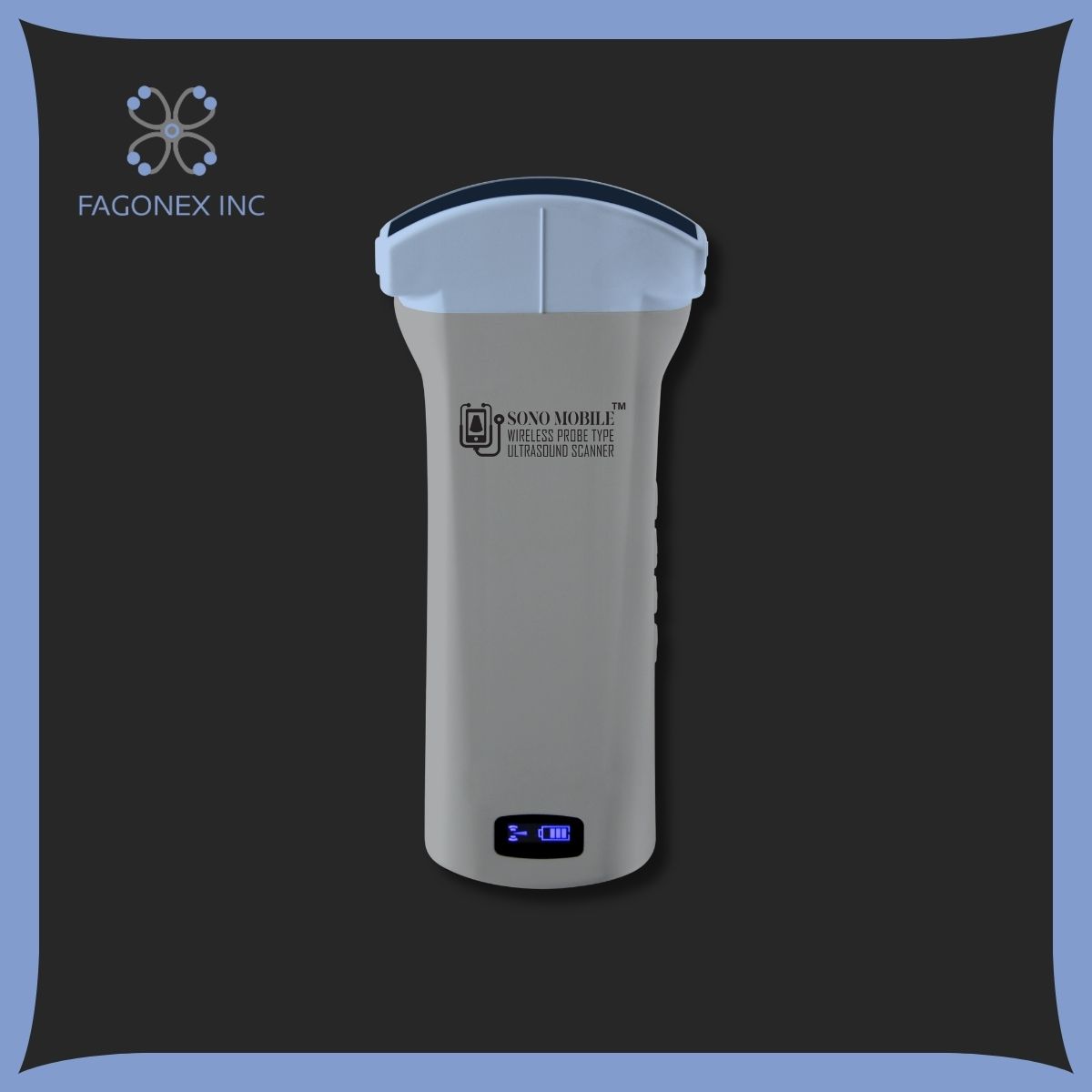
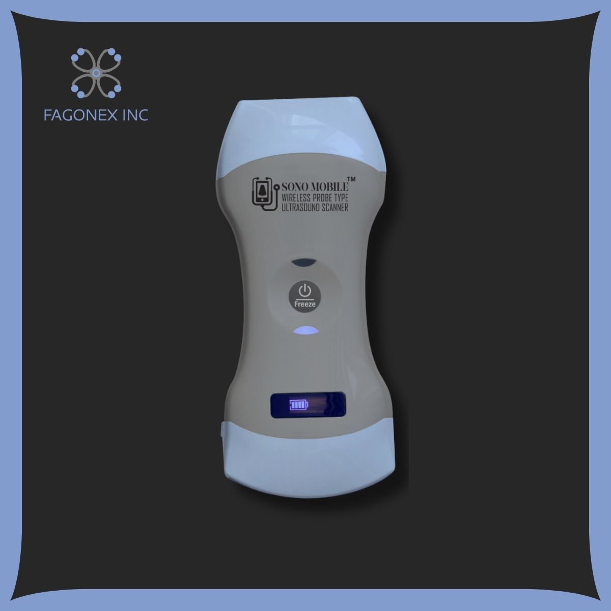
With the advent of portable devices like Sono Mobile™️CT61, pelvic ultrasounds are now more accessible than ever. Clinicians can perform scans at the point of care, in clinics, mobile health units, or even during home visits; offering diagnostic quality once limited to hospitals and large imaging centers.

Types: Transabdominal vs Transvaginal
There are two primary types of pelvic ultrasound: transabdominal and transvaginal. Each has specific clinical advantages and patient considerations.
Transabdominal Ultrasound
This is the more familiar external approach. The probe (transducer) is placed on the lower abdomen after applying a water-based gel. A full bladder is typically required, as it pushes pelvic organs into view and serves as an acoustic window. It offers a broad overview of the pelvic structures and is non-invasive and generally comfortable.
Ideal For:
General Gynecologic Evaluation
Pelvic ultrasound plays a central role in general gynecologic evaluations, offering non-invasive visualization of the uterus, ovaries, endometrium, and adnexa. It helps identify common conditions such as uterine fibroids, ovarian cysts, endometrial polyps, and polycystic ovary syndrome (PCOS). For women experiencing irregular bleeding, chronic pelvic pain, or abnormal menstrual cycles, this scan provides real-time insight for accurate diagnosis and treatment planning. Devices like Sono Mobile™️ CT61 allow physicians and OB-GYNs to perform these assessments in outpatient settings, women’s health clinics, or mobile care units, making high-resolution gynecologic ultrasound accessible anytime, anywhere; without delays or hospital referrals.
Early Pregnancy Scans
An early pregnancy scan between 6 to 12 weeks helps confirm fetal development, detect the gestational sac, yolk sac, and fetal heartbeat, and measure crown-rump length (CRL) to establish gestational age. It also rules out complications like ectopic pregnancy or missed miscarriage. Pelvic ultrasound is essential for reassuring parents and guiding next steps in prenatal care. Using a portable ultrasound system like Sono Mobile™️, clinicians can offer early scans in both clinic and home settings; empowering midwives, fertility specialists, and family physicians to provide first-trime
💧 Related Application: What Is a Bladder Scan?
While pelvic ultrasound examines reproductive organs, a bladder scan specifically assesses urine volume and retention. It’s quick, painless, and widely used in urology and emergency care.
ster care with professional accuracy and immediate results.
Patients Who Prefer an External Method
Many patients; especially those who are not sexually active or are uncomfortable with internal exams, prefer the external transabdominal ultrasound method. This non-invasive approach allows for effective pelvic imaging without the need for internal probe insertion, making it a suitable choice for young adults, adolescents, and individuals with personal or cultural sensitivities. While it requires a full bladder for optimal viewing, it still provides valuable insight into uterine size, ovarian structure, and early pregnancy status. Sono Mobile™️C6, with its high-quality abdominal imaging, offers a respectful and patient-centered solution for pelvic evaluation via external scanning.

Transvaginal Ultrasound
This approach involves inserting a slim, specially-designed probe into the vagina to obtain high-resolution images. It provides greater clarity and detail, especially useful for examining the endometrium, ovaries, and early fetal structures in early pregnancy. It requires informed consent and is generally well tolerated with minimal discomfort.
Ideal For:
- Early pregnancy complications
- Infertility assessments
- Detailed views of ovarian or uterine pathology
With Sono Mobile™️CT61, both methods are supported by adaptive imaging presets and multiple probe configurations. This flexibility ensures that providers can choose the most effective approach for each patient scenario, whether in an outpatient clinic or emergency setting.
📚 Inspired by Pelvic Ultrasound?
Interested in performing scans like these yourself? Explore our complete guide on How to Become an Ultrasound Technician — training paths, certifications, and what it takes to succeed.
Conditions Diagnosed with Pelvic Ultrasound
A pelvic ultrasound is essential for diagnosing and monitoring a wide range of reproductive, urological, and abdominal conditions. Its high diagnostic accuracy and non-invasive nature make it a first-line imaging modality in many clinical protocols.
Gynecologic Conditions:
- Uterine Fibroids: Benign muscular growths in the uterus that may cause pain, bleeding, or fertility issues.
- Ovarian Cysts: Fluid-filled sacs that are usually benign but need monitoring, especially if symptomatic or complex.
- Endometriosis: Although not directly visible, signs such as ovarian endometriomas or adhesions may suggest this condition.
- Polycystic Ovary Syndrome (PCOS): Characterized by multiple small follicles on the ovary with hormonal imbalances.
- Pelvic Inflammatory Disease (PID): Infection-related swelling and fluid accumulation in reproductive organs.
- Ectopic Pregnancy: A potentially life-threatening condition where the embryo implants outside the uterus.
- Miscarriage: Assessing fetal heartbeat and viability in early pregnancy.
- IUD Localization: Ensuring proper placement of intrauterine contraceptive devices.
Urological and Male Pelvic Conditions:
- Bladder Wall Abnormalities: Including thickening, tumors, or diverticula.
- Post-Void Residual Volume: Evaluated in cases of urinary retention or incomplete emptying.
- Prostate Assessment: Though less common than rectal ultrasound, pelvic ultrasound can detect prostate enlargement or lesions.
With Sono Mobile™️CT61, these conditions can be evaluated in real-time, making it a valuable tool for urgent care, family medicine, and rural health programs where immediate answers are critical for management.
Step-by-Step Guide to the Procedure
Understanding how a pelvic ultrasound is performed helps patients feel more informed and comfortable. Here’s a typical workflow for both scan types:
1. Preparation
- Transabdominal: Drink 3–4 glasses of water 1 hour before the exam; avoid urinating to keep the bladder full.
- Transvaginal: No specific preparation needed; the bladder should be empty for better imaging.
2. Positioning
- The patient lies on an exam table with the lower abdomen or pelvis exposed.
- For transabdominal scans, gel is applied to the abdomen.
- For transvaginal scans, the probe is covered with a protective sheath and lubricated.
3. Image Acquisition
- The probe is gently moved across the abdomen or inserted into the vagina.
- Real-time images are displayed on the ultrasound screen.
- The provider may ask the patient to slightly reposition or hold their breath briefly for better angles.
4. Measurements & Documentation
- Key measurements like uterine length, endometrial thickness, ovarian size, or CRL (in pregnancy) are taken.
- Still images or video loops are stored for diagnostic review or reports.
5. Completion
- The procedure typically lasts 10–20 minutes.
- There is no downtime; patients can resume normal activity immediately after.
Using Sono Mobile™️CT61, this process becomes even more streamlined. The device’s intuitive interface and multi-probe functionality allow clinicians to conduct the full procedure at the bedside, even in environments without access to electricity or fixed imaging infrastructure.

Post-Scan Expectations and FAQs
After the pelvic scan, patients can expect a quick return to normal life. In most cases, there are no side effects or recovery time needed.
What Happens After the Scan?
- The healthcare provider may review the images immediately and discuss findings.
- In some cases, the images are stored for review by a radiologist or specialist.
- If abnormalities are found, follow-up imaging or blood tests may be ordered.
Why Choose Sono Mobile™️ for Pelvic Ultrasound?
Sono Mobile™️ is designed with both portability and performance in mind, making it an ideal tool for pelvic imaging in diverse care settings; from hospital rooms and outpatient clinics to home visits and mobile outreach.
Key Features:
- High-Resolution Imaging: Capture detailed views of soft tissue, organs, and pathology.
- Multi-Probe Capabilities: Switch between transabdominal and transvaginal modes with ease.
- Wireless and Battery-Operated: Perform scans anytime, without needing a dedicated exam room or outlet.
- Lightweight and wireless Design: Perfect for midwives, OB-GYNs, and emergency responders.
- Smart Device Compatibility: View and share images via tablet, phone, or laptop system.
With Sono Mobile™️ CT61, you no longer have to wait days for diagnostic clarity. Provide real-time care, boost patient satisfaction, and elevate your clinical impact, wherever you practice.
Conclusion
A pelvic ultrasound is one of the most essential tools in diagnostic medicine, particularly in gynecology, reproductive health, and pelvic emergency care. Whether you’re evaluating unexplained pelvic pain, ruling out an ectopic pregnancy, or monitoring chronic conditions like PCOS, ultrasound offers a safe, accurate, and non-invasive solution.
Understanding the types of pelvic ultrasound; transabdominal vs transvaginal, and their applications empowers both patients and healthcare providers. And with innovations like Sono Mobile™️CT61, high-quality imaging is no longer limited to major hospitals or imaging centers. It’s now available at the point of care, helping you make faster, better-informed decisions.
Ready to Bring Pelvic Imaging to the Point of Care?
Don’t let access limit your diagnostic power. Sono Mobile™️ is the trusted solution for pelvic ultrasound in clinics, hospitals, emergency settings, and community health programs.
Whether you’re a general practitioner, OB-GYN, or urgent care provider, Fagonex helps you elevate patient care with the latest in mobile ultrasound technology.
Fagonex; Redefining access to care, one scan at a time.
Bring High-Quality Pelvic Imaging to Every Care Setting – Act Today
Whether you’re a gynecologist, family physician, midwife, or urgent care provider, having access to reliable, handheld ultrasound is no longer optional; it’s essential. With rising demand for on-site pelvic ultrasound, particularly for early pregnancy scans, gynecologic evaluations, and pelvic emergencies, you need a solution that is as mobile and efficient as your practice.
Sono Mobile™️ is the ultimate tool for delivering diagnostic excellence at the point of care. Its multi-probe system, high-resolution imaging, and seamless tablet/phone or laptop integration empower you to make faster, more accurate clinical decisions. From busy clinics to rural communities, from home visits to emergency rooms, Sono Mobile™️ brings flexibility, precision, and patient trust directly into your hands.
Don’t let outdated systems or access barriers delay your diagnosis or treatment planning. With Fagonex and Sono Mobile™️, you get the confidence of world-class technology backed by local support and training.
Contact Fagonex now for a personalized demo, pricing information, or to speak with an ultrasound expert.
Equip your practice. Empower your patients. Expand your impact; with Sono Mobile™️.
Fagonex – Advancing care, wherever you are.