Breast cancer screening and diagnostic evaluation rely mainly on two imaging methods: breast ultrasound and mammogram. These modalities differ fundamentally; ultrasound uses sound waves, while mammograms use low-dose X-rays, to offer complementary views of breast tissue. Understanding their strengths, limitations, and ideal use cases can empower women and clinicians to make informed decisions. This article compares breast ultrasound vs mammogram to aid in breast cancer screening and breast imaging strategies for women over 35 and those at higher risk.
Breast Ultrasound vs Mammogram
Breast ultrasound vs mammogram involves comparing two key imaging methods used in breast cancer screening and diagnosis. A mammogram uses low-dose X-rays and is the standard for routine screening, especially effective at detecting microcalcifications in women over 40. In contrast, a breast ultrasound uses sound waves to produce real-time images and is ideal for evaluating dense breast tissue, palpable lumps, or guiding biopsies. Ultrasound is radiation-free and preferred for younger or pregnant women, while mammograms are more sensitive to early-stage cancers. Both modalities are often used together for comprehensive breast evaluation.
What Is a Breast Ultrasound?
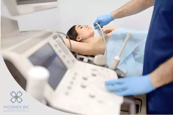
Breast ultrasound is a non-invasive imaging technique that uses high-frequency sound waves to create real-time images of breast tissue. It’s especially useful in evaluating palpable lumps, guiding biopsies, and further characterizing abnormalities first seen on mammogram or physical exam.
How It Works
- A transducer sends sound waves into the breast; reflected waves are captured and transformed into detailed grayscale images.
- It can distinguish between solid masses (which warrant further investigation) and benign fluid-filled cysts.
- No radiation is involved, making it safe for pregnant women and repeat examinations.
Advantages
- Highly sensitive in detecting lesions in dense breast tissue, which is common in younger women.
- Efficient for real-time evaluation; ultrasound-guided biopsy is minimally invasive and precise.
- Ideal for supplementary screening following inconclusive mammogram or for women with implants.
Limitations
- Less sensitive than mammography in detecting microcalcifications; a key early sign of ductal carcinoma in situ (DCIS).
- Operator dependency: image quality and interpretation depend on the sonographer’s skill.
- Not typically used as a standalone screening tool for women over 40 unless in conjunction with mammogram or high risk.
Role of Sono Mobile™️ in Modern Breast Imaging
Portable ultrasound technology is revolutionizing the landscape of breast cancer screening; and Sono Mobile™️ is at the forefront of this transformation. Designed by Fagonex Cooperation Inc., Sono Mobile™️ devices offer exceptional mobility, precision, and clarity, empowering healthcare providers to deliver high-quality breast imaging directly at the point of care.
Whether deployed in urban hospitals, rural clinics, private practices, or mobile screening units, Sono Mobile™️ enables healthcare professionals to perform breast ultrasound exams with confidence. This flexibility is critical in expanding access to diagnostic services for women in underserved or remote areas who may not have access to traditional mammography centers.
With high-resolution ultrasound imaging, Sono Mobile™️ devices provide detailed visualization of breast tissue, allowing clinicians to accurately differentiate between cystic and solid lesions. This capability is particularly valuable in younger women or those with dense breast tissue, where mammograms may miss smaller or hidden abnormalities. Early detection and characterization of suspicious areas lead to faster clinical decisions and timely referrals for biopsy or follow-up, when necessary.
Additionally, Sono Mobile™️ ultrasound systems are radiation-free, making them an ideal option for frequent breast examinations, especially for women under 40, pregnant patients, or individuals at higher risk for breast conditions. The intuitive interface and powerful measurement tools of Sono Mobile™️ streamline workflows and reduce diagnostic uncertainty, improving the patient experience and clinician efficiency.
By integrating cutting-edge breast ultrasound technology into compact and portable devices, Sono Mobile™️ bridges the gap between accessibility and advanced care. Its role in breast cancer screening strategies; whether as a stand-alone modality or a complementary tool to mammography, continues to grow, supporting earlier diagnosis and improved outcomes for women worldwide.
What Is a Mammogram?
A mammogram is a specialized breast X-ray and remains the cornerstone of routine breast cancer screening, recommended biennially or annually for women aged 40 or older.
How It Works
- The breast is gently compressed between plates; low-dose X-rays produce detailed images showing densities, masses, and microcalcifications.
- Both two-dimensional (2D) and three-dimensional (3D or tomosynthesis) imaging are used to enhance detection accuracy.
Advantages
- Superior at identifying microcalcifications, which can indicate early-stage cancers before palpable mass forms.
- Standardized and widely accepted in global screening guidelines.
- 3D tomosynthesis improves detection rates and reduces false positives, particularly in dense breasts.
Limitations
- In women with dense breast tissue, x-ray sensitivity is reduced, and additional imaging is often required.
- Exposure to low-dose radiation; though minimal, is still a consideration.
- Compression can cause discomfort and sometimes anxiety.
💡 Explore Related Imaging Insight
Curious how fetal brain development is monitored using ultrasound? Discover the full clinical significance of head circumference measurement in our dedicated guide:
HC Ultrasound Meaning.
Comparison Table: Ultrasound vs Mammogram
| Aspect | Ultrasound | Mammogram |
|---|---|---|
| Technique | Sound waves; no radiation | Low-dose X-ray; radiation involved |
| Ideal Use | Characterization of palpable lumps, cyst vs solid distinction, biopsy guidance, dense breasts | Routine screening, detection of microcalcifications, asymptomatic early detection |
| Strengths | No radiation; real-time imaging; portable (Sono Mobile™️ performance) | High sensitivity for calcifications; standardized screening; 3D tomosynthesis advantage |
| Limitations | Operator-dependent; limited detection of calcifications; not a stand-alone screen in asymptomatic older women | Less effective in dense tissue; radiation exposure; compression discomfort |
| Patient Use Cases | Women under 40; pregnant or lactating women; dense breast tissue; follow-up of abnormalities | Women 40+ for routine screening; complement to ultrasound findings |
When to Choose Each Method
Routine Screening (Asymptomatic Women)
- Mammogram is the gold standard for average-risk women ≥ 40 years old; typically performed every 1–2 years depending on local guidelines and patient risk profile.
- Breast ultrasound may be added for women with dense breasts or risk factors (family history, genetic predisposition) to improve detection sensitivity.
Younger or Pregnant Women
- Ultrasound is the preferred initial imaging method for women < 30 years, pregnant, or lactating; as it is safe and effective in excluding benign entities without exposing to radiation.
Palpable Lump or Abnormal Mammogram
- Ultrasound is used to further evaluate palpable masses, confirm cystic vs solid findings, and facilitate ultrasound-guided breast biopsy when necessary.
- In some cases, both imaging modalities and MRI may be combined for further assessment.
High-Risk Patients
- Women with BRCA mutations or strong family history may undergo annual mammogram plus supplemental ultrasound or MRI, especially if they have dense tissue.
Post-Surgical or Implant Evaluation
- Ultrasound evaluates for implant rupture, fluid collection, or scar tissue; used as a supplement or when mammogram images are inconclusive due to hardware artifact.
Breast Cancer Detection Rates and Limitations
Mammogram Detection Effectiveness
- Mammography demonstrates high sensitivity (around 85–90%) in fatty breasts, detecting early-stage cancers and microcalcifications.
- In dense breasts, sensitivity can decrease to 60–70%, resulting in false negatives.
- Recall rates for additional imaging can be 10–15%, with a false-positive biopsy or call-back rate around 5–10%.
Ultrasound Detection Effectiveness
- Ultrasound effectively identifies cysts versus solid lesions and detects cancers missed on mammogram, particularly in dense breasts.
- Sensitivity ranges between 70–95% depending on operator expertise. It cannot reliably visualize microcalcifications, which limits its standalone screening capacity for early-stage DCIS.
Complementarity and Workflow Integration
- Combined use of mammogram and breast ultrasound in dense breasts increases overall cancer detection by about 30%, though it also increases false positives.
- Workflow typically involves routine mammogram first, followed by targeted ultrasound based on findings or in high-risk scenarios.
- Portable devices like Sono Mobile™️ enable quick bedside imaging, instant lesion characterization, and faster diagnostic resolution; especially valuable in community clinics or follow-up visits.

Role of Sono Mobile™️ in Improving Breast Imaging Access
In the evolving field of breast cancer screening, accessibility to timely and accurate diagnostic tools is more crucial than ever. Traditional breast ultrasound machines, while effective, are often large, immobile, and require hospital-based infrastructure; making them less accessible in community clinics, primary care offices, or underserved rural settings. Enter Sono Mobile™️, a breakthrough solution developed by Fagonex Cooperation Inc., that’s redefining how and where breast imaging can take place.
Sono Mobile™️ is a handheld, portable ultrasound system that delivers hospital-grade imaging performance in a lightweight and compact design. Specifically engineered to meet the needs of point-of-care breast imaging, it is ideal for physicians, nurse practitioners, and sonographers working in non-hospital environments such as outreach programs, mobile screening units, telemedicine platforms, and disaster relief missions.
With its advanced image resolution, Sono Mobile™️ allows clinicians to clearly identify subtle differences in tissue composition, making it easier to detect and differentiate between benign cysts and solid masses. This is particularly beneficial for women under 40 and those with dense breast tissue, where mammograms may not provide sufficient detail. Moreover, the radiation-free nature of breast ultrasound makes Sono Mobile™️ a safer choice for repeat evaluations and for patients who require frequent monitoring due to elevated risk factors.
By bringing diagnostic-grade breast imaging to the bedside, exam room, or field clinic, Sono Mobile™️ empowers providers to offer breast cancer screening to populations previously beyond reach. This democratization of care is essential in achieving early detection, reducing disparities in outcomes, and supporting global efforts in women’s health. For healthcare professionals looking to expand their services or improve efficiency, Sono Mobile™️ offers a powerful, scalable solution tailored for today’s dynamic clinical environments.
Clinical Advantages
- Enables immediate ultrasound-guided breast biopsy, reducing diagnosis delays.
- Helps triage patients effectively, ensuring those with suspicious findings are referred promptly.
- Enhances comfort and convenience; no need for mammography compression for initial lump evaluation.
Operational Impact
- Reduces the burden on imaging centers by enabling early assessment in satellite clinics.
- Ensures seamless digital integration; images and measurements can be sent directly to PACS or EMR systems.
- Supports patient education: real-time review of images helps providers explain findings, options, and next steps.
Conclusion
In the ongoing debate of breast ultrasound vs mammogram for breast cancer screening and diagnosis, both modalities play unique and complementary roles:
- Mammogram remains the foundational screening tool for women over 40; excellent at detecting microcalcifications and early-stage disease.
- Breast ultrasound excels in evaluating palpable lumps, dense breast tissue, and guiding biopsies, but lacks calcification sensitivity.
Integrating both modalities, particularly for women at higher risk or with dense breasts, offers the most comprehensive strategy for breast cancer detection and personalized care. With portable ultrasound innovation like Sono Mobile™️, breast imaging is becoming more accessible, responsive, and patient-centered; empowering clinicians and women alike to achieve earlier detection and better outcomes.
If you’re a woman over 35, at higher risk of breast disease, or seeking more information on breast imaging options, consult your healthcare provider about combining mammogram and ultrasound screening; including the use of advanced, portable solutions like Sono Mobile™️, for the highest level of care.