
Musculoskeletal (MSK) ultrasound is a powerful, non-invasive imaging technique that has transformed the way clinicians evaluate and treat injuries related to muscles, tendons, ligaments, and joints. Using high-frequency sound waves, MSK ultrasound provides real-time visualization of internal soft tissue structures, making it an essential diagnostic tool in modern medicine.
With the emergence of high-quality portable systems like Sono Mobile™️, musculoskeletal imaging is more accessible than ever, supporting on-site assessments in clinics, emergency rooms, physiotherapy settings, and sports fields. At Fagonex, we are dedicated to advancing diagnostic capabilities by offering state-of-the-art MSK ultrasound solutions to healthcare professionals in orthopedics, physiotherapy, rheumatology, and sports medicine.
What Is MSK Ultrasound?
MSK ultrasound (musculoskeletal ultrasound) refers to a diagnostic procedure that uses sound waves to create visual images of muscles, joints, tendons, ligaments, and soft tissues. Unlike MRI or CT scans, this imaging technique can be performed dynamically, allowing practitioners to observe structures in motion; such as a tendon gliding during wrist flexion or fluid accumulation during knee flexion.
Through real-time scanning, clinicians can gain insights into inflammation, tears, impingements, and other subtle yet significant changes in soft tissue structures. The Sono Mobile™️ portable ultrasound device, known for its clarity and ease of use, is increasingly becoming the first choice for musculoskeletal assessments worldwide.
Diagnostic Applications
The diagnostic versatility of MSK ultrasound makes it an invaluable tool for assessing a wide variety of injuries and disorders, especially in settings requiring quick and accurate information. It can be used to:
- Diagnose tendon injuries such as rotator cuff tears or Achilles tendinopathy
Musculoskeletal ultrasound is a first-line tool for evaluating tendon injuries. For example, rotator cuff tears; common in athletes and aging adults, can be clearly visualized with MSK imaging, detecting both partial and full-thickness tears. Similarly, Achilles tendinopathy, which often develops from overuse or improper footwear, shows characteristic thickening and hypoechoic regions on ultrasound. With Sono Mobile™️, clinicians can perform real-time, high-resolution tendon assessments right at the point of care, making ultrasound for injuries an indispensable asset in sports medicine and orthopedic settings.
- Evaluate ligament injuries including ACL sprains or medial collateral ligament tears
Ligament injuries often present with pain, swelling, and instability, particularly in high-impact or twisting injuries. MSK ultrasound offers a dynamic, radiation-free approach to visualizing ligament integrity, identifying sprains, partial ruptures, and even chronic laxity. In the case of ACL sprains or medial collateral ligament (MCL) tears, diagnostic ultrasound allows clinicians to observe real-time joint movement and detect fluid buildup or fiber discontinuity. Using portable systems like Sono Mobile™️, healthcare professionals can make accurate, on-the-spot assessments that support immediate treatment decisions.
- Examine joint effusions, synovial thickening, or early rheumatoid arthritis changes
Joint inflammation and effusion are hallmark signs of underlying pathology such as arthritis, gout, or infection. Musculoskeletal ultrasound is highly sensitive for detecting small amounts of intra-articular fluid, synovial hypertrophy, and early erosive changes, especially in conditions like rheumatoid arthritis. These findings are often missed in early-stage disease by X-rays. With real-time MSK imaging, clinicians using Sono Mobile™️ can monitor disease progression or response to therapy, making diagnostic ultrasound an ideal tool in rheumatology and internal medicine practices.
- Visualize muscle tears, hematomas, or contusions in real-time
Muscle injuries can range from minor contusions to severe ruptures and hematomas. Ultrasound for injuries provides unmatched resolution of superficial muscle fibers, detecting disruptions, fluid collections, and dynamic changes during movement. For athletes and trauma patients, musculoskeletal ultrasound offers a fast, bedside solution to differentiate between strains and more serious tears. With portable technology like Sono Mobile™️, clinicians can visualize and document muscle pathology in seconds, aiding in quicker rehabilitation planning and minimizing time away from sports or work.
- Detect nerve entrapments like carpal tunnel syndrome or ulnar neuropathy
Entrapment neuropathies are commonly misdiagnosed without imaging, leading to prolonged discomfort or inappropriate treatment. MSK imaging allows direct visualization of nerve swelling, compression points, and surrounding anatomical structures. In conditions like carpal tunnel syndrome, the median nerve can be tracked as it passes through the wrist, showing characteristic enlargement and decreased mobility. Similarly, ulnar neuropathy at the elbow is easily assessed. Using diagnostic ultrasound with tools like Sono Mobile™️, providers can identify the root cause of symptoms and improve long-term patient outcomes.
- Assess bursitis, cysts, and other soft tissue masses
When patients present with localized swelling or pain, it’s often due to fluid-filled structures such as bursae or cysts. Musculoskeletal ultrasound helps distinguish between solid masses, abscesses, and benign fluid collections. For instance, in bursitis, ultrasound reveals bursal distension, wall thickening, and surrounding inflammation. Ganglion cysts or Baker’s cysts are also clearly delineated. With Sono Mobile™️, clinicians can perform detailed soft tissue evaluations at the bedside or in outpatient settings, making ultrasound for injuries and abnormalities a go-to diagnostic option in general practice and specialty clinics.
This real-time MSK imaging capability is crucial for quick diagnoses and timely intervention, especially in sports medicine and physiotherapy environments where accurate, on-the-spot decisions are needed.
💡 Also worth reading: HC Ultrasound Meaning – Understand how head circumference is measured in fetal scans and why it matters for healthy development.
Advantages Over Other Imaging
MSK ultrasound has several distinct advantages over traditional imaging modalities like MRI or CT:
- Radiation-Free: Since it uses sound waves, it poses no radiation risk, making it ideal for repeat imaging or use in children and pregnant patients.
- Real-Time Assessment: Structures can be evaluated while in motion, a key feature for identifying dynamic joint and tendon abnormalities.
- Highly Portable: Devices like Sono Mobile™️ are compact and user-friendly, enabling point-of-care diagnostics in diverse environments.
- Cost-Effective: Ultrasound is generally more affordable than MRI or CT, particularly for ongoing monitoring of injuries or therapy progression.
- Superior for Soft Tissues: While MRI is excellent for deeper joint and bone imaging, ultrasound often offers better resolution of superficial tendons, muscles, and ligaments.
For healthcare providers focused on efficiency, accuracy, and safety, MSK ultrasound has become the gold standard for diagnostic ultrasound of the musculoskeletal system.
How It’s Done
The procedure for MSK ultrasound is straightforward, safe, and generally well-tolerated:
- Preparation: The patient is positioned to expose the affected area, often seated or lying down.
- Gel Application: A water-based gel is applied to ensure optimal contact and eliminate air between the probe and skin.
- Scanning: The clinician moves the transducer over the area, capturing live images.
- Dynamic Testing: The patient may be asked to perform specific movements to evaluate joint behavior, tendon gliding, or impingement during activity.
- Interpretation: Images are analyzed on the spot, and the results are often discussed with the patient immediately.
The procedure typically takes 15–30 minutes and requires no downtime or recovery.
Diagnosing Injuries
Ultrasound for injuries offers unique advantages over static imaging because of its ability to detect subtle, movement-based abnormalities. This is especially relevant for:
Shoulder Pain: Detection of rotator cuff tears or bursitis
Shoulder pain is a prevalent musculoskeletal complaint, often caused by conditions such as rotator cuff tears or subacromial-subdeltoid bursitis. Musculoskeletal ultrasound plays a critical role in diagnosing these issues with high sensitivity. Using MSK imaging, clinicians can evaluate tendon integrity, identify fluid accumulation, and assess inflammation in real time. This is especially valuable for patients with limited mobility or those who cannot undergo MRI. Sono Mobile™️ devices provide immediate bedside evaluation, allowing for dynamic assessment of shoulder structures during movement. With diagnostic ultrasound, clinicians can better differentiate between impingement syndromes and soft tissue abnormalities, leading to faster, targeted treatments.
Elbow Injuries: Diagnosis of tennis elbow or medial epicondylitis
Elbow pain is frequently associated with overuse injuries such as lateral epicondylitis (tennis elbow) and medial epicondylitis (golfer’s elbow). These conditions involve inflammation and microtears in the tendons attaching to the lateral or medial epicondyle of the humerus. Musculoskeletal ultrasound is a non-invasive, efficient imaging modality for diagnosing tendon thickening, calcifications, and increased vascularity. With ultrasound for injuries, physicians can also assess adjacent structures like bursae and ligaments. Sono Mobile™️ enhances point-of-care decision-making by offering real-time diagnostic ultrasound for quick and accurate evaluation, making it especially beneficial for orthopedic, sports medicine, and physiotherapy specialists.
Wrist & Hand Conditions: Carpal tunnel syndrome, tendonitis, and joint effusion detection
The wrist and hand are prone to repetitive stress injuries, making them common sites for carpal tunnel syndrome, tenosynovitis, and joint effusions. MSK imaging provides detailed visualization of the median nerve within the carpal tunnel, detecting nerve swelling and compression with excellent sensitivity. Additionally, diagnostic ultrasound identifies inflamed tendons and fluid in tendon sheaths or joints, often missed in routine exams. With portable systems like Sono Mobile™️, healthcare providers can conduct focused, dynamic ultrasound for injuries at the point of care. This aids in early detection, reduces the need for costly imaging, and ensures precise clinical intervention in wrist and hand disorders.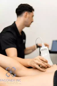
Knee Injuries: Identifying effusions, patellar tracking issues, or meniscal abnormalities
Knee pain can stem from various sources, including joint effusions, patellar maltracking, or meniscal injuries. While MRI is often used, musculoskeletal ultrasound is gaining prominence as a first-line imaging method for evaluating superficial knee structures. MSK imaging allows visualization of effusions, quadriceps or patellar tendon disorders, and signs of synovial inflammation. With diagnostic ultrasound, clinicians can perform real-time assessments during flexion and extension, aiding in the diagnosis of dynamic patellar instability. Sono Mobile™️ provides quick and accurate knee evaluations without radiation exposure, making it a go-to tool in physiotherapy, sports medicine, and orthopedics for efficient joint imaging and injury monitoring.
Ankle & Foot Problems: Achilles tendon rupture, plantar fasciitis, or ligament instability
Foot and ankle injuries are common in both athletes and the general population, with conditions such as Achilles tendon ruptures, plantar fasciitis, and ligament instability requiring precise diagnosis. Musculoskeletal ultrasound is ideal for visualizing soft tissue abnormalities in the lower extremities, including tears, inflammation, and fluid accumulation. MSK imaging can dynamically assess the Achilles tendon under stress, detect plantar fascia thickening, and evaluate lateral ligaments for sprains or chronic laxity. With the portability of Sono Mobile™️, healthcare providers can perform immediate ultrasound for injuries in clinics, emergency rooms, or field settings. This fast, cost-effective diagnostic ultrasound tool enhances treatment accuracy and speeds up recovery planning.
We encourage our readers to also explore our article Ultrasound for Diagnosing Musculoskeletal Injuries for a deeper understanding of how ultrasound is applied across various clinical scenarios.
Why Choose Ultrasound?
Healthcare professionals often ask why ultrasound should be chosen over more traditional imaging like MRI or CT. Here’s why:
- Instant Feedback: Clinicians and patients receive immediate results.
- Dynamic Assessments: No other imaging allows real-time movement analysis like MSK ultrasound.
- Safe and Repeatable: Especially important for athletes undergoing multiple assessments or chronic pain patients who need ongoing monitoring.
- Improved Patient Outcomes: Accurate diagnosis leads to quicker treatment plans and faster recovery.
- Guided Therapy: Used to precisely guide injections, aspirations, or biopsies, increasing both efficacy and patient safety.
To compare this modality further, visit our educational guide on the Difference Between Ultrasound and CT Scan.
Case Studies
Real-world examples illustrate how effective MSK ultrasound can be:
Case 1 – Rotator Cuff Injury in a Swimmer
A 29-year-old professional swimmer reported shoulder pain. Using Sono Mobile™️, a partial-thickness tear of the supraspinatus tendon was detected. Immediate referral to physiotherapy and activity modification led to full recovery in six weeks.
Case 2 – Chronic Ankle Pain in a Runner
A 40-year-old runner experienced recurring pain during training. Ultrasound showed peroneal tendon subluxation not detected on previous X-rays. Early diagnosis allowed surgical planning that avoided further tendon damage.
Case 3 – Office Worker with Wrist Pain
A 52-year-old with wrist discomfort was misdiagnosed with arthritis. MSK ultrasound revealed tenosynovitis due to repetitive strain, and conservative treatment resolved symptoms in four weeks.
These examples highlight the necessity and power of ultrasound for injuries, especially in active individuals and professionals requiring accurate diagnosis for rapid recovery.
Real-World Examples in Specialty Practices
The scope of MSK ultrasound extends far beyond orthopedics. Here’s how it’s used across different specialties:
Rheumatology: Evaluate joint effusions, synovitis, and erosion in patients with rheumatoid arthritis
In rheumatology, early and accurate diagnosis of conditions like rheumatoid arthritis is essential to prevent long-term joint damage. Musculoskeletal ultrasound allows direct visualization of joint effusions, synovial hypertrophy, and early erosive changes that may not appear on X-rays. With MSK imaging, rheumatologists can track inflammation levels, guide joint aspirations, and monitor response to treatment over time. Diagnostic ultrasound using portable systems like Sono Mobile™️ offers dynamic, real-time feedback during clinical exams, making it a valuable tool in both outpatient and bedside settings for timely and precise joint imaging.
Physiotherapy: Monitor muscle healing and guide rehabilitation in soft tissue injuries
Physiotherapists increasingly rely on musculoskeletal ultrasound to monitor tissue regeneration and healing after injuries such as muscle tears, tendonitis, or ligament sprains. With MSK imaging, therapists can visually assess the progress of soft tissue injuries, ensuring that rehabilitation exercises are targeted and safe. Ultrasound for injuries also allows for early detection of complications like scar tissue or hematomas. The portability of Sono Mobile™️ brings diagnostic ultrasound directly into the therapy setting, enabling real-time feedback during movement and improving outcomes through personalized, data-driven recovery programs.
Sports Medicine: On-site evaluation of injuries during training or competition
In sports medicine, time is critical when dealing with athletic injuries. Musculoskeletal ultrasound offers a rapid, accurate solution for evaluating injuries on-site, whether during training or live competition. MSK imaging enables immediate assessment of sprains, muscle tears, or joint effusions, allowing for quick decisions on whether an athlete can safely return to play. Sono Mobile™️ systems are designed for field use, making diagnostic ultrasound accessible in gyms, sidelines, and clinics. By offering handheld ultrasound for injuries, sports medicine professionals can deliver timely care and minimize downtime for athletes.
Orthopedics: Assess surgical outcomes, implant positions, and guide procedures
Orthopedic specialists benefit greatly from musculoskeletal ultrasound in both surgical planning and post-operative care. MSK imaging can verify proper implant positioning, assess for complications like effusions or tendon damage, and guide minimally invasive procedures such as joint aspirations or injections. Diagnostic ultrasound is a powerful, non-radiative tool for monitoring healing, evaluating grafts, and checking soft tissue around surgical sites. With Sono Mobile™️, orthopedic surgeons can perform dynamic, bedside joint imaging for more accurate, efficient follow-up care. This enhances clinical confidence and helps patients return to function faster.
Chiropractic and Pain Clinics: Identify sources of inflammation or nerve impingement for treatment planning
Chiropractors and pain management specialists use musculoskeletal ultrasound to evaluate soft tissue structures and identify underlying causes of chronic pain. MSK imaging is ideal for detecting nerve impingements, bursitis, tendonitis, or localized inflammation that may be causing musculoskeletal discomfort. By using real-time diagnostic ultrasound, clinicians can pinpoint treatment areas and guide therapies such as dry needling, trigger point injections, or adjustments. With compact tools like Sono Mobile™️, ultrasound for injuries and nerve assessment becomes an efficient addition to the clinical workflow, improving diagnostic accuracy and treatment outcomes in chiropractic and pain clinic settings.
Visit our specialty pages to learn more about how MSK ultrasound fits into your specific area of practice:
Conclusion
Musculoskeletal ultrasound is no longer an emerging option; it’s a cornerstone of modern musculoskeletal care. From diagnosing acute injuries in athletes to managing chronic joint conditions, MSK imaging offers real-time, safe, and accurate visualization of the human body’s complex structures.
At Fagonex, our mission is to empower healthcare providers with cutting-edge tools like Sono Mobile™️ that bring portability, precision, and performance together. Whether you’re working in orthopedics, sports medicine, or physical therapy, MSK ultrasound will enhance your diagnostic capabilities, improve patient satisfaction, and elevate your practice.
Explore more on our website or contact us to discover how our mobile ultrasound solutions can revolutionize your diagnostic workflow.