
What Is OFD in Ultrasound?
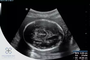
In prenatal ultrasound evaluations, OFD, or occipitofrontal diameter, is a critical fetal head measurement that helps healthcare professionals assess a baby’s development inside the womb. The OFD represents the distance between the occiput (back of the fetal skull) and the forehead (frontal bone). It is measured on an axial plane of the fetal head, usually taken during the second trimester or later.
Using high-resolution, portable devices like the Sono Mobile™️ CT61, sonographers and physicians can acquire accurate and real-time measurements of fetal biometry, including the OFD, during routine pregnancy scans. This parameter offers insight into fetal growth patterns, brain development, head shape, and potential abnormalities.
While less commonly discussed than BPD (biparietal diameter) or HC (head circumference), OFD is equally essential in providing a three-dimensional understanding of the fetal head. It assists not only in identifying normal growth trajectories but also in recognizing issues like brachycephaly (shortened OFD) or dolichocephaly (elongated OFD).
Role of OFD in Fetal Head Measurements
Why Measure OFD?
The occipitofrontal diameter is an essential component of comprehensive fetal head evaluation. When combined with BPD and HC( HC Ultrasound Meaning), OFD provides a more accurate assessment of cranial shape and volume. These measurements help determine if the fetus is growing appropriately for its gestational age and whether there are signs of cranial shape distortion.
In particular, the shape of the head is influenced by genetic, positional, and sometimes pathological factors. Measuring OFD helps in:
- Diagnosing head shape abnormalities
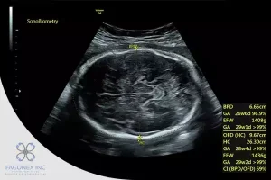
- Calculating the cephalic index (CI), which is BPD/OFD × 100
- Supporting gestational age estimation
- Monitoring fetal growth trends over time
- Flagging signs of intrauterine constraint or pathology
How Is OFD Measured?
The OFD is typically measured in the transventricular plane, an axial slice through the fetal head where the midline falx, cavum septum pellucidum, and thalami are visible. The measurement is taken from the outer edge of the occipital bone to the outer edge of the frontal bone, using calipers aligned parallel to the midline.
With the portability and precision of devices like the Sono Mobile™️ CT61, clinicians can easily capture and analyze OFD during routine checkups, even in remote or point-of-care environments, ensuring that care is accessible and consistent.
Normal OFD Ranges by Week
Understanding the normal range for OFD values is essential in distinguishing between normal growth patterns and potential abnormalities. Below is a table showing average OFD values by gestational age:
Normal OFD Ranges (Occipitofrontal Diameter)
| Gestational Age (Weeks) | Mean OFD (mm) | Normal Range (mm) |
| 14 weeks | 27 mm | 25–29 mm |
| 16 weeks | 32 mm | 30–34 mm |
| 18 weeks | 37 mm | 35–39 mm |
| 20 weeks | 42 mm | 40–44 mm |
| 22 weeks | 47 mm | 45–49 mm |
| 24 weeks | 52 mm | 50–54 mm |
| 26 weeks | 57 mm | 55–59 mm |
| 28 weeks | 62 mm | 60–64 mm |
| 30 weeks | 67 mm | 65–69 mm |
| 32 weeks | 71 mm | 69–73 mm |
| 34 weeks | 74 mm | 72–76 mm |
| 36 weeks | 77 mm | 75–79 mm |
| 38 weeks | 80 mm | 78–82 mm |
| 40 weeks | 83 mm | 81–85 mm |
These ranges are approximate and may slightly vary between institutions and ultrasound protocols.
An OFD that falls below or above the expected range could prompt further investigation. For instance:
- Low OFD + normal BPD → Suggests brachycephaly
- High OFD + low BPD → Suggests dolichocephaly
- Low OFD and BPD → May indicate growth restriction
🧬 Related Topic: Pelvic Ultrasound
OFD is just one part of prenatal imaging. Want to understand broader reproductive scans? Read Pelvic Ultrasound: What Is It to explore how pelvic anatomy is assessed during early pregnancy and beyond.
How OFD Complements Other Metrics
OFD alone is helpful, but its true value is realized when interpreted alongside other fetal head parameters, especially in assessing fetal growth and development holistically.
OFD and BPD (Biparietal Diameter)
BPD measures the width of the fetal head, while OFD measures the length. When combined, these values help determine the cephalic index (CI):
CI = (BPD / OFD) × 100
- Normal CI: 74–83%
- High CI (>85%) suggests brachycephaly (short skull)
- Low CI (<70%) suggests dolichocephaly (long, narrow skull)
OFD and HC (Head Circumference)
While OFD and BPD are linear measurements, head circumference (HC) offers a perimeter-based value, providing insight into overall head size. HC is the most commonly used parameter for estimating fetal age and brain volume. OFD supports the calculation by offering more data on head elongation or compression, which might affect HC values.
OFD and Skull Shape Abnormalities
Monitoring OFD helps detect abnormal cranial shapes:
| Condition | OFD Trend | BPD Trend | Cephalic Index |
| Brachycephaly | Decreased | Increased | High |
| Dolichocephaly | Increased | Decreased | Low |
Both conditions are usually benign, but in some cases, positional molding or premature suture fusion (craniosynostosis) might be considered, especially when values are extreme or associated with other anomalies.
Use of Sono Mobile™️ CT61 in Biometry
The Sono Mobile™️ CT61 is a next-generation portable ultrasound system designed for high-performance point-of-care imaging. It offers clinicians fast and accurate access to fetal biometric measurements, including BPD, FL, and AC. Engineered for both precision and convenience, this device features a dual-probe design; combining both convex and endovaginal probes in a single unit. With the push of a button, users can seamlessly switch between modes, dramatically increasing the mobility, efficiency, and clinical readiness of the system in diverse obstetric settings.
Its high-resolution imaging, lightweight ergonomic design, and intuitive touchscreen interface make the Sono Mobile™️ CT61 ideal for obstetricians, gynecologists, radiologists, and sonographers working in hospitals, women’s health clinics, fertility centers, and even mobile ultrasound services. Preloaded fetal biometry presets and caliper guidance reduce manual steps and minimize user error, making it a powerful solution for both routine prenatal care and advanced fetal assessments.
Final Thoughts: Why OFD Matters
The occipitofrontal diameter (OFD) is a powerful yet often underappreciated measurement in fetal biometry. It adds value not only in understanding the size of the fetal head but also its shape, aiding in the diagnosis of positional or developmental abnormalities.
When integrated with other fetal metrics, OFD enhances the reliability of gestational age estimation, provides clues about fetal wellbeing, and supports clinical decision-making.
Thanks to innovative imaging solutions like the Sono Mobile™️ CT61, measuring OFD has become more accessible, precise, and efficient; empowering healthcare professionals and reassuring expectant parents with detailed insight into fetal development.
Learn More or Request a Demo
At Fagonex Cooperation Inc., we are committed to advancing mobile and point-of-care ultrasound technologies. Our flagship product, Sono Mobile™️ CT61, offers best-in-class features for obstetric imaging, including OFD and comprehensive fetal measurements.
Whether you’re a healthcare provider, medical student, or clinic/hospital seeking clarity, our team is here to guide you.
📞 Contact us today to learn more, or schedule a live demo of the Sono Mobile™️ CT61.
🔍 Fascinated by OFD Measurements?
Understanding fetal head dimensions is just one part of the journey. Learn How to Become an Ultrasound Technician and gain the skills to perform and interpret vital scans like OFD with confidence.