
Prenatal ultrasound is a cornerstone of obstetric care. With precise imaging, healthcare providers can monitor fetal development, guide clinical decision‑making, and support the emotional well‑being of pregnant individuals. In this comprehensive guide, tailored to OB/GYN patients, caregivers, and expecting parents, we’ll demystify the timeline, purpose, and interpretation of pregnancy ultrasound both for routine use and in special cases.
What Is the Role of Ultrasound in Pregnancy?
A Safe, Non‑Invasive Pregnancy Ultrasound Modality
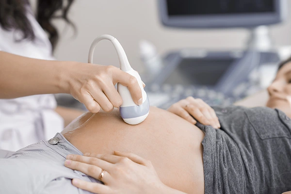
Ultrasounds use high-frequency sound waves to produce real‑time visual images of the fetus, placenta, and uterus. They are painless, radiation-free, and widely accepted as safe throughout pregnancy. The signal is interpreted by specially trained sonographers or OB/GYNs.
The Impact of Modern Tools
Advanced technology, such as the Sono Mobile™️ CT61, enables high-resolution imaging even in outpatient or mobile care settings. Physicians and clinics adopting this portable ultrasound device benefit from enhanced pregnancy ultrasound capabilities; improving diagnostic clarity while boosting patient convenience.
Three Core Functions of Ultrasound
- Dating and Viability: Confirms pregnancy and estimates gestational age.
- Anomaly Detection: Screens for anatomical or genetic irregularities.
- Growth Monitoring: Tracks fetal development and amniotic fluid balance.
First Trimester: Dating and Viability Scans
Why Early Scanning Matters
Early trimester scans, typically between 6–12 weeks, are crucial. They confirm an intrauterine pregnancy, assess viability (heartbeat), and establish an accurate due date.
Gestational Age Estimation
Measurement of Crown–Rump Length (CRL) around 6–10 weeks provides the most precise dating, significantly influencing the entire ultrasound schedule and determining delivery timing.
Detecting Early Challenges
Sometimes, early scans detect issues such as missed or threatened miscarriage, molar pregnancy, or ectopic gestation; situations that require immediate clinical attention.
Nuchal Translucency and Risk Assessment
Between 11–14 weeks, a combined scan includes measuring nuchal translucency (NT) thickness, used alongside blood tests to evaluate risk for chromosomal conditions like Down syndrome.
Expectation of a First‑Trimester Scan
Transvaginal approach offers superior detail before week 10.
A transvaginal pregnancy ultrasound is preferred before week 10 for high-resolution imaging. It helps detect early gestational structures and provides clearer fetal development details during the first trimester scan.
After week 10, transabdominal scanning may suffice.
After 10 weeks, a transabdominal ultrasound generally offers sufficient detail. This method is non-invasive and effective for ongoing pregnancy monitoring in second and third trimester scans using devices like Sono Mobile™ CT61.
Heartbeat typically visible by days 6–7 post-conception.
By day 6 or 7 post-conception, a fetal heartbeat can often be visualized with early pregnancy ultrasound. Confirming viability early on reassures both patients and caregivers about pregnancy progress.
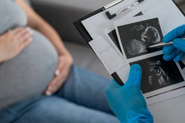
Sonographer records gestational sac, yolk sac, CRL, heartbeat, and location.
During a first trimester ultrasound, the sonographer documents key markers: gestational sac, yolk sac, crown-rump length (CRL), fetal heartbeat, and intrauterine location; essential for assessing healthy pregnancy development.
Second Trimester: Anomaly and Growth Checks

The Anatomy Scan: Routine Mid-Pregnancy Evaluation
Between 18–22 weeks, the detailed anomaly scan evaluates vital fetal structures, brain, heart, spine, face, kidneys, limbs; and genetic markers like limb length and nuchal skin fold.
Placenta, Amniotic Fluid & Position
Checks include placental attachment and maturity, amniotic fluid volume (AFI), and uterine artery flow to assess risk of preeclampsia or growth restrictions.
Growth Assessment
Estimated fetal weight (EFW) is calculated, compared with gestational age, and plotted on growth charts. Early detection of macrosomia (large baby) or intrauterine growth restriction (IUGR) helps guide optimal delivery planning.
The Role of Sono Mobile™️ CT61
This device supports high-resolution mid-pregnancy imaging. Its portability means critical scans can be delivered conveniently; especially useful in remote or resource-limited clinics.
Multiple Pregnancies & Anomalies
In twins or multiples, both fetuses are individually examined. Chorionicity and amnionicity are established before 14 weeks; subsequent scans monitor twin-specific complications like twin‑to‑twin transfusion.
🧬 Related Topic:
We suggest you read this article. How Many Ultrasounds Are Needed During Pregnancy
Third Trimester and Late Pregnancy Scans
Why Late Term Ultrasound Matters
From approximately 28 weeks onward, trimester scans grow more targeted:
- Third Trimester Late Scans (34–38 weeks) monitor fetal growth trajectory, movement, presentation, and fluid volume.
- Used to plan delivery strategies in pregnancies with diabetes, hypertension, or multiple gestation.
Monitoring Fetal Growth
By measuring head circumference, femur length, and abdominal circumference, clinicians can track growth velocity and identify deviations suggestive of macrosomia or IUGR.
Determining Fetal Presentation
Positioning matters: identifying breech or transverse lie 4–6 weeks before delivery informs decisions around vaginal birth vs. cesarean section.
Assessing Placental & Fluid Health
Placental maturation, calcifications, and amniotic fluid index (AFI) are tracked; critical in diagnosing oligohydramnios or polyhydramnios, both risk factors for preterm labor and complications.
When Additional Scans Are Needed
Conditions like hypertension, gestational diabetes, or decreased fetal movements may warrant more frequent pregnancy ultrasound monitoring. The Sono Mobile™️ CT61 offers efficient, onsite scans to allay concern and update care plans.
Interpreting Results and Common Concerns
Understanding Standard Findings
A typical reading includes:
Gestational Age
Gestational age is estimated using pregnancy ultrasound, typically based on crown-rump length (CRL) in the first trimester. Accurate dating helps guide prenatal care, growth milestones, and delivery timing.
Fetal Measurements vs. Percentile for Age
Ultrasound tracks fetal measurements like head, abdomen, and femur length, comparing them to standard growth percentiles. Falling below or above average may indicate growth restrictions or macrosomia risks.
Amniotic Fluid Index
The amniotic fluid index (AFI) is measured via transabdominal ultrasound to assess fetal well-being. Low AFI (oligohydramnios) or high AFI (polyhydramnios) can signal complications requiring close monitoring.
Placental Grade and Location
A pregnancy ultrasound evaluates placental grade and location, identifying issues like placenta previa or calcification. Placental health is vital for nutrient delivery and fetal oxygenation throughout gestation.
Fetal Heart and Breathing Motion
Ultrasound assesses fetal heart rate and breathing motion, key indicators of fetal viability. Regular rhythm and visible diaphragmatic motion suggest a healthy, developing baby during routine trimester scans.
Biophysical Profile Score
The biophysical profile (BPP) combines ultrasound and non-stress test to evaluate fetal movements, tone, breathing, and fluid levels. A high score reflects good fetal health, guiding decisions in late pregnancy.
Normal findings are reassuring; minor variations are common and rarely alarming.
Abnormal Results & Next Steps
Parents may hear terms like:
- Anomaly Detected: Requires referral to a perinatologist.
- IUGR or SGA (Small for Gestational Age): May need serial scans and growth monitoring.
- Oligohydramnios/Polyhydramnios: Requires closer fluid surveillance.
- Placenta Previa/Accreta: Influences delivery planning and monitoring.
Follow‑Up Strategies
- Additional scans scheduled per condition severity.
- Non‑stress tests (NST) or biophysical profiles (BPP) assess fetal well‑being.
- If necessary, hospital transfers and consultations with maternal‑fetal medicine specialists.
Special Case: Multiple Pregnancies
Frequent scans account for selective growth, spacing, and shared versus separate chorionic/amniotic sacs. Monitoring identifies preterm labor risk or discordance.
Sensitive Results Conversations
Good providers tailor delivery of unexpected or concerning findings with empathy, provide emotional support, and outline necessary tests or referrals to guide parents confidently forward.
Best Practices for an Ultrasound‑Friendly Pregnancy Experience
Prepare for the Scan
- Early scans: a moderately full bladder improves image clarity.
- Second/third trimester: Drink fluids beforehand, but don’t overfill.
- Wear comfortable, loose clothing; consider bringing your partner or support person.
Ask Questions
- “What is the estimated gestational age?”
- “Are there any structural concerns?”
- “How is the placenta situated?”
- “Can you show us the heart, spine, limbs?”
Follow‑Up Action Steps
- Note any recommended re‑scans or referrals.
- Track growth metrics if they’re shared.
- Contact your provider with questions or concerns about symptoms or fetal movement.
The Role of Sono Mobile™️ CT61 in Modern Prenatal Care
Portability Meets Picture Quality
The Sono Mobile™️ CT61 combines portability with a powerful imaging suite. Expectant families benefit from bedside clarity and rapid scanning; even outside traditional hospital settings.
Advantages for Clinics and OB/GYN Practices
- Supports outpatient and community‑based clinics, remote deployment, and home‑visit scenarios.
- Enables real‑time diagnostic capabilities without sacrificing image quality.
- Ideal for follow‑ups (e.g., then‑and‑now comparison scans) and specialized use like amniotic fluid measurement, growth monitoring, or follow‑up of anomalies.
Designing an Effective Prenatal Ultrasound Schedule
A general prenatal ultrasound roadmap, incorporating Sono Mobile™️ CT61:
| Trimester | Weeks | Purpose | Scan Focus |
| First | 6–12 | Dating, viability, NT screening | CRL, heartbeat, NT, gestational sac |
| Mid‑pregnancy | 18–22 | Anomaly and growth check | Anatomy, EFW, placenta, fluid, umbilical flow |
| Late pregnancy | 32–36* | Growth, presentation, planning | Biometry, AFI, positioning |
| Special cases | As needed | IUGR, multiples, placenta concerns | Tracking dynamics, BPP, NST, Dopplers |
* timing may vary; some obstetricians incorporate a 28‑week check or repeat scans at 34 weeks depending on medical history and risk factors.
Key Takeaways for Pregnant Individuals and Caregivers
- Ultrasound is safe and central to modern prenatal care.
- Trimester scans follow diagnostic milestones; from confirming life and dating to anomaly checks and late‑term planning.
- The Sono Mobile™️ CT61 supports accurate imaging with accessible and portable convenience.
- Understanding scan results empowers informed decisions and timely interventions.
Follow‑up plans; whether re‑scans, specialist referrals, or fetal‑well‑being tests, stem from your results.