
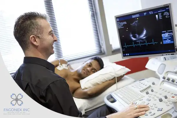
Ultrasound has become a cornerstone in the field of cardiology, offering non-invasive and highly accurate insights into heart function, structure, and blood flow dynamics. Known as cardiac ultrasound or echocardiography, this imaging modality has transformed the diagnosis, monitoring, and treatment of cardiovascular diseases.
This article delves into the types, benefits, and future trends of cardiac ultrasound, emphasizing its significance in modern cardiology.
 ejection fraction, ventricular function, and fluid status, guiding treatment adjustments in heart failure patients.
Monitoring Chronic Conditions
For patients with chronic diseases like hypertension or atrial fibrillation, regular ultrasounds track disease progression and treatment efficacy.
Point-of-Care Applications
POCUS devices enable clinicians to perform cardiac evaluations during emergencies, offering quick assessments of conditions like pulmonary embolism or acute heart failure.
The portability of modern ultrasound machines ensures seamless integration into various care settings, from ICUs to outpatient clinics.
Introduction to Cardiac Ultrasound
Cardiac ultrasound, commonly referred to as echocardiography, uses high-frequency sound waves to create detailed images of the heart and its surrounding structures. It is widely utilized to assess heart function, detect abnormalities, and guide treatment decisions. Unlike other imaging modalities, echocardiography is non-invasive, safe, and capable of providing real-time visuals. From portable devices to advanced systems like point-of-care ultrasound (POCUS), cardiac ultrasound is pivotal in both clinical and emergency settings.Types of Cardiac Ultrasound
The versatility of cardiac ultrasound is evident in its various types, each catering to specific diagnostic needs.Transthoracic Echocardiography (TTE)
This is the most common form of cardiac ultrasound. A transducer is placed on the chest to capture detailed images of the heart. TTE is used to evaluate heart size, structure, and motion.Transesophageal Echocardiography (TEE)
In TEE, a specialized probe is inserted into the esophagus to obtain clearer images of the heart, especially the posterior structures. It is often used in cases requiring high-resolution imaging.Doppler Echocardiography
This technique measures the speed and direction of blood flow within the heart and blood vessels. Doppler imaging is crucial for diagnosing conditions like valve regurgitation or stenosis.Stress Echocardiography
Performed during or after physical stress or pharmacological stimulation, this type evaluates how well the heart functions under increased workload. It is commonly used to diagnose coronary artery disease.3D and 4D Echocardiography
Advancements in technology have enabled three-dimensional imaging of the heart, providing comprehensive views of its anatomy. 4D echocardiography adds real-time motion, making it ideal for surgical planning and intraoperative assessments.Benefits of Ultrasound in Cardiology
Non-Invasive and Safe Echocardiography eliminates the need for invasive procedures, reducing patient risk. Its non-ionizing nature ensures safety, even for repeated use. Real-Time Imaging The ability to observe heart function in real time provides immediate insights, aiding rapid decision-making in critical situations. Wide Applicability From routine checkups to emergency care, cardiac ultrasound serves diverse purposes, including the diagnosis of congenital defects, cardiomyopathies, and valve diseases. Portability and Accessibility Modern portable ultrasound machines and POCUS devices like Sono Mobile™️ have expanded the reach of echocardiography, enabling bedside and remote care. These tools are particularly useful in underserved areas and emergency situations.How Ultrasound Assists in Diagnosing Heart Conditions
Heart Valve Disorders Echocardiography evaluates the function and structure of heart valves, detecting stenosis, regurgitation, and prolapse. Doppler imaging is particularly effective in assessing blood flow patterns. Congenital Heart Defects For infants and children, echocardiography identifies structural abnormalities such as atrial septal defects (ASD) and ventricular septal defects (VSD). Cardiomyopathies Cardiac ultrasound assesses the thickness, motion, and size of heart walls, aiding in the diagnosis of hypertrophic, dilated, or restrictive cardiomyopathies. Pericardial Effusion Echocardiography detects fluid accumulation around the heart, helping diagnose pericarditis or cardiac tamponade. Coronary Artery Disease Stress echocardiography evaluates myocardial perfusion and wall motion abnormalities, identifying areas of ischemia.Role of Ultrasound in Monitoring Cardiac Patients
Post-Surgical Evaluation Echocardiography monitors heart function and structural changes following procedures such as valve replacement or coronary artery bypass grafting. Heart Failure Management Sono Mobile™️ Cardiac ultrasound assesses ejection fraction, ventricular function, and fluid status, guiding treatment adjustments in heart failure patients.
Monitoring Chronic Conditions
For patients with chronic diseases like hypertension or atrial fibrillation, regular ultrasounds track disease progression and treatment efficacy.
Point-of-Care Applications
POCUS devices enable clinicians to perform cardiac evaluations during emergencies, offering quick assessments of conditions like pulmonary embolism or acute heart failure.
The portability of modern ultrasound machines ensures seamless integration into various care settings, from ICUs to outpatient clinics.