
BPD Ultrasound Meaning: What Does BPD Stand For?
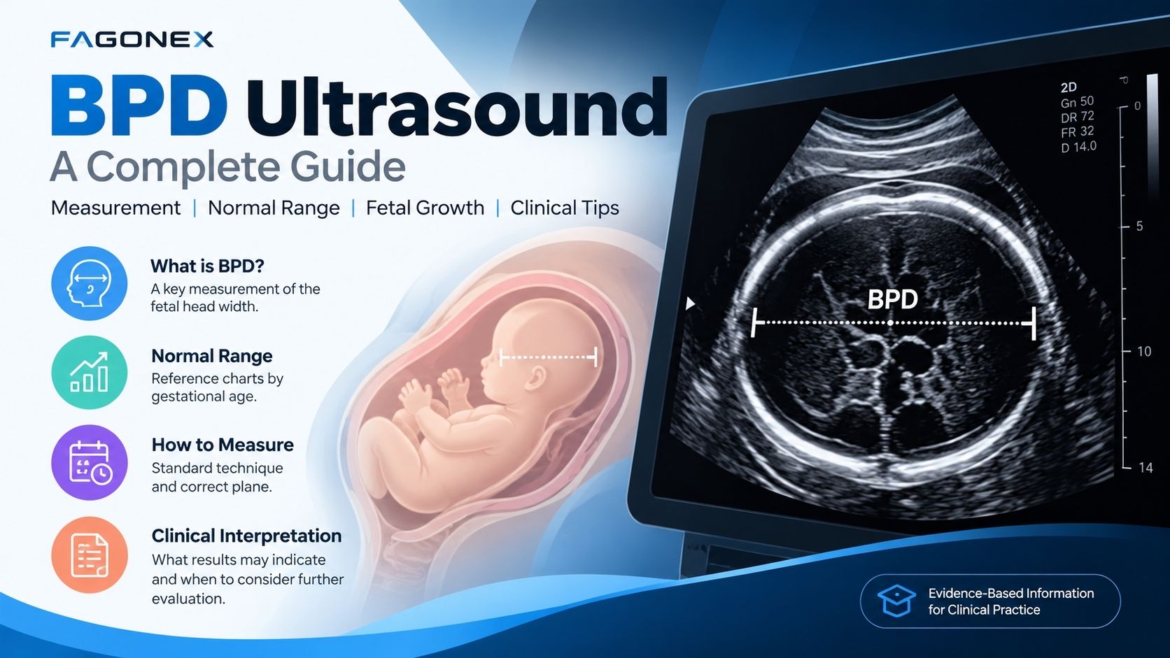
BPD (biparietal diameter) is an ultrasound measurement of the width of a baby’s head during pregnancy. It is one of several measurements clinicians use to assess fetal size and growth during pregnancy.
BPD is obtained on a standardized transverse view of the fetal head. It is interpreted alongside head circumference (HC), abdominal circumference (AC), femur length (FL), gestational age, and the overall clinical picture—not as a stand-alone indicator of fetal health.
BPD may be measured during the second-trimester anatomy scan (often around 18–22 weeks) and during other indicated fetal growth examinations. The timing and choice of measurements depend on the clinical assessment.
Why Is BPD Important in Fetal Growth?
BPD is part of standard fetal biometry. Its value is most useful when the measurement technique is consistent and the result is compared with an appropriate reference chart.
Gestational age assessment
BPD can contribute to pregnancy dating, although early ultrasound dating is generally preferred when available.
Growth monitoring
Serial fetal measurements can help clinicians assess growth patterns over time.
Head size assessment
BPD and HC provide information about fetal head dimensions; they do not diagnose brain abnormalities by themselves.
Clinical planning
Biometry contributes to overall obstetric assessment together with other findings and clinical factors.

BPD Ultrasound Normal Range: Measurements by Pregnancy Week
The following values are approximate illustrative ranges reproduced from the existing FAGONEX article, not a validated clinical reference chart. Actual percentiles depend on the selected growth standard, dating accuracy, and measurement technique.
| Gestational age | Approximate BPD range |
|---|---|
| 13 weeks | 22 – 25 mm |
| 14 weeks | 25 – 28 mm |
| 15 weeks | 28 – 31 mm |
| 16 weeks | 31 – 34 mm |
| 17 weeks | 34 – 37 mm |
| 18 weeks | 37 – 41 mm |
| 19 weeks | 41 – 45 mm |
| 20 weeks | 45 – 49 mm |
| 21 weeks | 49 – 53 mm |
| 22 weeks | 53 – 56 mm |
| 23 weeks | 56 – 60 mm |
| 24 weeks | 60 – 63 mm |
| 25 weeks | 63 – 66 mm |
| 26 weeks | 66 – 69 mm |
| 27 weeks | 69 – 72 mm |
| 28 weeks | 72 – 75 mm |
| 29 weeks | 75 – 78 mm |
| 30 weeks | 78 – 81 mm |
| 31 weeks | 81 – 83 mm |
| 32 weeks | 83 – 86 mm |
| 33 weeks | 86 – 88 mm |
| 34 weeks | 88 – 91 mm |
| 35 weeks | 91 – 93 mm |
| 36 weeks | 93 – 95 mm |
| 37 weeks | 95 – 97 mm |
| 38 weeks | 97 – 99 mm |
| 39 weeks | 99 – 100 mm |
| 40 weeks | 100 – 102 mm |
What Is BPD at 19 or 20 Weeks?
People often look for a single BPD value at 19 or 20 weeks. The table above shows illustrative ranges of 41–45 mm at 19 weeks and 45–49 mm at 20 weeks, but these figures are not validated clinical percentiles. An individual measurement must be assessed against a validated reference standard for the confirmed gestational age.
Important: Do not use this table to diagnose abnormal fetal growth or make clinical decisions. A qualified healthcare professional should interpret BPD using validated gestational-age-specific charts and other fetal biometric findings.
How Is Biparietal Diameter Measured on Ultrasound?
Accurate BPD measurement requires the correct imaging plane, appropriate caliper placement, and a trained operator.
1. Obtain the correct plane
Acquire a symmetrical transverse view of the fetal head showing relevant landmarks, including the thalami and cavum septi pellucidi, with the midline appropriately aligned.
2. Place the calipers consistently
Depending on the reference standard, BPD may be measured outer-to-inner or outer-to-outer. Use the convention specified by the clinical protocol and reference chart.
3. Review image quality
Check fetal position, image symmetry, and landmark visibility. Repeat the measurement if the plane is oblique or the landmarks are inadequate.
4. Interpret with other biometry
Review BPD alongside HC, AC, FL, clinical history, and any other recommended measurements.
Common measurement challenges
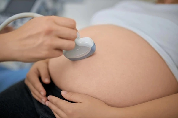
- Fetal movement or position: may make a suitable transverse plane difficult to obtain.
- Head shape: can influence BPD; head circumference may be particularly useful when shape is atypical.
- Operator technique: inconsistent landmarks or caliper placement can affect results.
Frequently Asked Questions About BPD Ultrasound
What is the full form of BPD in pregnancy ultrasound?
BPD stands for biparietal diameter. It measures the transverse width of the fetal head on a standardized ultrasound plane and is one component of fetal biometry.
What does a low BPD measurement mean?
A BPD measurement below an expected percentile does not establish a diagnosis on its own. Dating, head shape, other biometric measurements, and clinical findings must be reviewed by the care team.
What does a high BPD measurement mean?
A higher-than-expected BPD can have several explanations, including normal variation or differences in fetal head shape. It should be assessed using appropriate reference charts and the full ultrasound examination.
Is BPD enough to estimate fetal weight?
No. Estimated fetal weight typically uses a combination of biometric measurements, such as HC, AC, and FL, according to the selected formula and clinical protocol.
At what stage of pregnancy is BPD measured?
BPD is commonly included in second-trimester fetal biometry and may also be obtained at later growth scans when clinically indicated.
Can handheld ultrasound be used for fetal biometry?
Some portable ultrasound systems may support fetal imaging and measurements, depending on their specifications, regulatory indications, imaging quality, and operator training. Device suitability should be verified for the intended clinical use.
Explore Portable Ultrasound for Clinical Practice
FAGONEX offers Sono Mobile® wireless handheld ultrasound solutions, including the convex-probe Sono Mobile® C6. Explore device specifications and contact the team to discuss suitability for your intended application. Product capabilities should not be taken as a claim of validated fetal biometry performance.
Key Takeaways: Understanding BPD Ultrasound Results
- BPD stands for biparietal diameter, a standard measurement of fetal head width.
- BPD is interpreted alongside other measurements, not in isolation.
- Measurement technique and the selected reference chart affect interpretation.
- Questions about an individual ultrasound result should be discussed with a qualified prenatal care professional.
Medical information notice: This article is for general education and is not a substitute for professional medical advice, diagnosis, or treatment.
Medical References and Clinical Guidelines
The following clinical guidelines and fetal growth standards provide additional information about biparietal diameter (BPD), fetal biometry, ultrasound measurement techniques, and interpretation.
- ISUOG Practice Guidelines: Ultrasound Assessment of Fetal Biometry and Growth (2019).
- AIUM Practice Parameter for the Performance of Standard Diagnostic Obstetric Ultrasound (2024).
- ISUOG Practice Guidelines: Performance of the Routine Mid-Trimester Fetal Ultrasound Scan (2022).
- INTERGROWTH-21st: International Fetal Growth Standards and BPD Charts .