
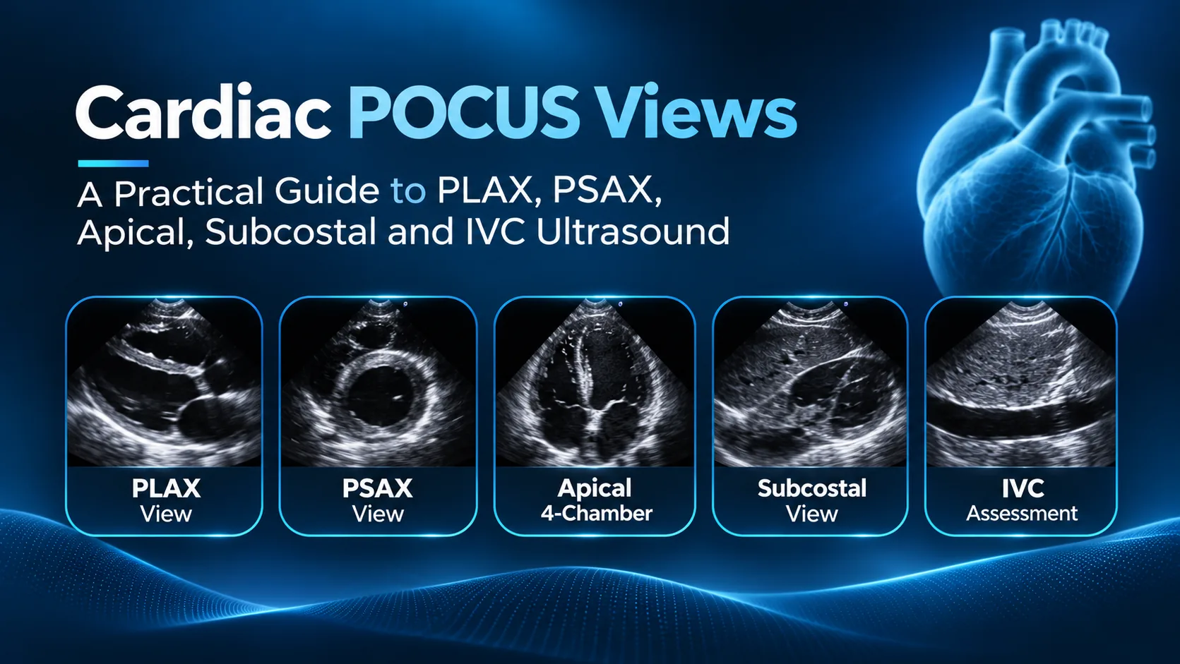
FAGONEX Clinical Education · Cardiac Point-of-Care Ultrasound
Cardiac POCUS Views: A Practical Guide to PLAX, PSAX, Apical & Subcostal Ultrasound
Learn the standard cardiac POCUS views, where to position the probe, which anatomical landmarks to identify, and how to recognize selected bedside findings. Explore five real cardiac ultrasound demonstrations acquired using Sono Mobile® CL64.
Clinical fundamentals
What Are the Standard Cardiac POCUS Views?
Cardiac point-of-care ultrasound (POCUS), often termed focused cardiac ultrasound (FoCUS), is a limited bedside examination used by appropriately trained clinicians to address specific clinical questions. Unlike a comprehensive transthoracic echocardiogram, FoCUS is not intended to provide a complete structural, Doppler, or quantitative cardiac assessment. The examination is interpreted alongside the patient's symptoms, physical examination, vital signs, and other investigations. [1, 2]
Parasternal windows
PLAX and PSAX provide complementary views of ventricular anatomy, cardiac motion, and selected pericardial findings.
Apical window
The apical four-chamber view helps compare right and left ventricular size and observe atrioventricular structures.
Subcostal window
May provide a useful alternative when parasternal or apical windows are difficult and supports focused pericardial assessment.
IVC window
Shows inferior vena cava size and respiratory behavior, interpreted cautiously in the broader hemodynamic context.
For broader clinical applications, visit the FAGONEX Cardiology specialty page or explore our POCUS resource hub.
View 01 · Parasternal
Parasternal Long-Axis (PLAX) Ultrasound View
The parasternal long-axis view is typically obtained near the left sternal border, often around the third or fourth intercostal space, with the cardiac probe marker directed toward the patient's right shoulder under the commonly taught cardiac convention. Orientation conventions vary by equipment and training program; always confirm the displayed image orientation.
Key landmarks: right ventricular anterior wall/outflow region, interventricular septum, left ventricle, mitral valve, left atrium, aortic root, and aortic valve. The descending thoracic aorta may help distinguish pericardial from left pleural fluid in this window. [2]
Real PLAX ultrasound demonstration · Sono Mobile® CL64
What to look for
- Gross left ventricular contraction and chamber dimensions.
- Pericardial fluid, when visible, with attention to its anatomical location.
- Mitral and aortic valve motion as a qualitative observation, not a complete valve assessment.
Common acquisition problem
An oblique or off-axis image can misrepresent chamber size. Make small rotational, tilting, and sliding adjustments to obtain a true long-axis plane.
View 02 · Parasternal
Parasternal Short-Axis (PSAX) Ultrasound View
The parasternal short-axis view is generally acquired by rotating the transducer approximately 90 degrees from a well-aligned PLAX position, with the marker oriented toward the patient's left shoulder in a commonly used cardiac convention. Tilting through different levels reveals the aortic valve, mitral valve, papillary muscles, and left ventricular apex.
At the mid-ventricular papillary-muscle level, the left ventricle normally appears approximately circular. A flattened interventricular septum may produce a D-shaped left ventricle, a finding associated with right ventricular pressure or volume overload in the appropriate context; it is not diagnostic of pulmonary embolism or any single condition by itself. [2, 3]
PSAX and D-shaped septum demonstration · Sono Mobile®
What to look for
- Left ventricular shape and qualitative systolic contraction.
- Septal flattening and possible right ventricular loading patterns.
- Regional motion differences, recognizing the limitations of a single plane.
Common acquisition problem
A short-axis image obtained at the wrong level or off-axis can create misleading ventricular geometry. Identify the anatomical level before interpreting septal shape.
View 03 · Apical
Apical Four-Chamber (A4C) Cardiac Ultrasound View
The apical four-chamber view is acquired near the cardiac apex, often with the patient in a left lateral position when feasible. Direct the ultrasound beam toward the base of the heart and optimize the plane to display both atria and both ventricles. The exact probe-marker direction depends on the imaging convention and equipment setup.
This view supports qualitative comparison of right and left ventricular size, observation of global cardiac motion, and identification of the mitral and tricuspid valves. Avoid apical foreshortening, which can distort ventricular length and make functional interpretation less reliable. [2, 3]
Apical four-chamber demonstration · Sono Mobile® CL64
What to look for
- Relative right and left ventricular dimensions.
- Gross global systolic function.
- Visible atrioventricular anatomy and chamber relationships.
Common acquisition problem
Foreshortening occurs when the scan plane misses the true apex. Reposition the probe to maximize ventricular length and avoid drawing conclusions from a truncated chamber.
View 04 · Subcostal
Subcostal Four-Chamber View and Pericardial Effusion
The subcostal four-chamber view is often obtained just below the xiphoid process, using the liver as an acoustic window while directing the beam toward the patient's left shoulder. It can be especially useful when other cardiac windows are technically limited.
The subcostal window is frequently used to look for pericardial effusion. An anechoic space around the heart may indicate fluid, but image quality, distribution, and alternative explanations must be considered. The presence of pericardial fluid alone does not establish cardiac tamponade; tamponade is a clinical and hemodynamic diagnosis requiring urgent integrated assessment. [1, 2]
Subcostal cardiac and pericardial fluid demonstration · Sono Mobile® CL64
What to look for
- Pericardial fluid surrounding cardiac structures.
- Gross cardiac activity in a window less dependent on intercostal access.
- Findings requiring additional views and urgent clinical correlation.
Common acquisition problem
Excessive pressure, inadequate depth, or a poor liver window can obscure the heart. Adjust depth and angle while maintaining patient comfort.
View 05 · Subcostal venous assessment
Inferior Vena Cava (IVC) Ultrasound in Cardiac POCUS
The IVC ultrasound view is commonly obtained through a subcostal longitudinal window, displaying the inferior vena cava as it enters the right atrium. The clinician observes vessel caliber and changes with respiration while maintaining alignment with the vessel's long axis.
IVC diameter and respiratory variation can contribute to an assessment of right atrial pressure under appropriate conditions. However, the relationship is affected by respiratory effort, mechanical ventilation, intra-abdominal pressure, right-heart pathology, and other factors. IVC findings alone should not be used to diagnose dehydration, determine fluid responsiveness, or guide fluid therapy. [3]
IVC ultrasound demonstration · Sono Mobile® CL64
What to look for
- IVC continuity into the right atrium.
- Respiratory changes in IVC caliber.
- Possible off-axis or translation artifacts that affect interpretation.
Clinical connection
Cardiac and IVC windows may be incorporated into a broader RUSH examination for shock and hypotension, together with lung, abdominal, and vascular assessment.
At-a-glance reference
Cardiac POCUS Views: Quick Comparison
| View | Primary anatomy | Focused clinical use | Important limitation |
|---|---|---|---|
| PLAX | LV, LA, mitral and aortic structures | Gross LV function; selected pericardial findings | Off-axis chamber distortion |
| PSAX | LV cross-section, septum, RV relationship | Qualitative contraction; septal shape | Interpretation depends on imaging level |
| A4C | Both atria and ventricles | Chamber comparison; gross function | Apical foreshortening |
| Subcostal | Four chambers and pericardial space | Pericardial fluid; alternative cardiac window | Effusion does not alone diagnose tamponade |
| IVC | IVC and right atrial junction | Contextual venous assessment | Not a stand-alone fluid responsiveness test |
Image acquisition
How to Improve Cardiac POCUS Image Quality
- Start with the clinical question. Define whether you are evaluating gross ventricular function, pericardial fluid, or another focused finding.
- Choose the cardiac preset and appropriate depth. Ensure the relevant cardiac structures fit within the field of view.
- Confirm orientation. Probe-marker conventions and screen presentation may differ between devices and teaching systems.
- Optimize the acoustic window. Adjust patient position when safe, then slide, rotate, fan, or tilt the probe gradually.
- Use multiple complementary views. Do not make major clinical conclusions from a single poor-quality image.
- Document findings and limitations. Escalate to formal echocardiography or specialist review when indicated.
For a broader discussion of bedside imaging, see what point-of-care ultrasound is and how it is used across clinical specialties. For focused shock assessment, see our RUSH exam ultrasound guide.
From learning to bedside practice
Using Handheld Ultrasound for Cardiac POCUS
Portable ultrasound can support supervised training and focused bedside imaging when the system provides appropriate cardiac imaging capabilities and is used by a qualified clinician. The demonstrations above were recorded using Sono Mobile® CL64, a wireless dual-head handheld ultrasound system with a phased-array cardiac preset. Device selection should account for image quality, clinical requirements, operator training, and local regulatory requirements.
Learn more about cardiology ultrasound applications, explore POCUS education resources, or review the Sono Mobile® CL64 product specifications.
Explore Sono Mobile® CL64 for Cardiac POCUS
See the handheld ultrasound system featured in these five clinical demonstrations. Review its cardiac preset, imaging modes, connectivity, and device specifications, or contact FAGONEX to discuss a product demonstration.
Frequently asked questions
Cardiac POCUS Views: FAQs
What are the four standard cardiac POCUS views?
They are the parasternal long-axis (PLAX), parasternal short-axis (PSAX), apical four-chamber (A4C), and subcostal four-chamber views. IVC assessment is often added as a complementary subcostal venous view.
What is the difference between PLAX and PSAX?
PLAX displays the heart along its long axis, including the left ventricle, mitral valve, and aortic root. PSAX provides cross-sectional views at different levels, useful for evaluating left ventricular shape and qualitative motion.
What does a D-shaped left ventricle mean on PSAX?
Septal flattening can create a D-shaped left ventricular appearance and may reflect right ventricular pressure or volume overload. Interpretation depends on timing, other ultrasound findings, and the clinical context; it does not identify a single diagnosis.
Can a subcostal view identify pericardial effusion?
It may reveal fluid in the pericardial space, but further assessment is needed to determine significance. An effusion by itself does not establish tamponade.
Can IVC ultrasound determine whether a patient needs fluids?
No. IVC measurements are influenced by multiple physiologic and technical factors and should not be used alone to predict fluid responsiveness or direct treatment.
Does cardiac POCUS replace a full echocardiogram?
No. FoCUS answers selected bedside questions and complements clinical evaluation. Comprehensive echocardiography is needed when a complete structural, functional, Doppler, or quantitative assessment is indicated.
Evidence-based education
Scientific References & Clinical Guidelines
- Spencer KT, Kimura BJ, Korcarz CE, Pellikka PA, Rahko PS, Siegel RJ. Focused Cardiac Ultrasound: Recommendations from the American Society of Echocardiography. Journal of the American Society of Echocardiography. 2013;26(6):567–581. DOI / publication.
- British Society of Echocardiography. Guidance on standard adult transthoracic echocardiographic views and acquisition. Open-access professional resource. Open-access guideline.
- American Society of Echocardiography. Guidelines for the Echocardiographic Assessment of the Right Heart in Adults and Special Considerations in Pulmonary Hypertension. 2025. Guideline PDF.
Educational videos: FAGONEX / Sono Mobile®. The linked professional guidance is independent and does not imply endorsement of FAGONEX or its products. Last editorial review: November 2026.