
FAGONEX | Medical Ultrasound Education
Handheld Ultrasound in Medical Training: Benefits, Applications & POCUS Education
Handheld ultrasound is changing how medical students, residents, and practicing clinicians learn to connect anatomy with bedside assessment. Portable systems can bring supervised scanning into classrooms, simulation labs, and clinical training environments—helping learners develop practical point-of-care ultrasound (POCUS) skills.
This guide explains where handheld ultrasound fits into medical training, what an effective learning program looks like, and which device features matter when educators select equipment.
Why Handheld Ultrasound Matters in Medical Training
Ultrasound offers a dynamic view of anatomy that static diagrams cannot provide. During a supervised scan, learners can observe how organs, vessels, and soft tissues appear in real time, then relate those images to physical examination and clinical questions.
Traditional cart-based ultrasound systems remain important for comprehensive imaging and advanced instruction. Handheld devices complement them by making it easier to organize small-group practice, bedside demonstrations, and repeated skills sessions in suitable teaching settings.
For a broader overview of educational use cases and equipment considerations, visit the FAGONEX ultrasound in medical education page.
Benefits of Handheld Ultrasound for Medical Students
Real-Time Anatomy Visualization
Scanning can help students connect anatomical landmarks with live cross-sectional images, movement, and variation between individuals.
Hands-On Clinical Skills
Regular guided practice supports probe handling, image orientation, depth and gain adjustment, and recognition of common structures.
Portable Learning
Compact equipment can support demonstrations and practice in simulation rooms, classrooms, and approved clinical environments.
These advantages are educational opportunities, not automatic improvements in diagnostic accuracy. Outcomes depend on curriculum quality, instructor expertise, practice time, and competency assessment.
How Handheld Ultrasound Supports POCUS Education
Point-of-care ultrasound (POCUS) uses focused ultrasound examinations to answer specific clinical questions at or near the patient's bedside. Training should teach both image acquisition and the limits of a focused examination.
Bedside Scanning and Clinical Decision-Making
Under appropriate supervision, learners can practice translating a clinical question into a scanning protocol, identifying relevant anatomy, and recognizing when findings require confirmation or escalation. Interpretation should always be integrated with the patient's history, examination, and other clinical information.
Simulation, Workshops, and Supervised Practice
Skills stations and simulation-based exercises allow educators to teach probe positioning, common scanning windows, and troubleshooting before students apply those skills in clinical settings. Repeated feedback and documented assessment are more meaningful than device access alone.
A practical POCUS learning sequence
- Define the clinical question and learning objective.
- Review anatomy, safety, and examination limitations.
- Demonstrate the scanning protocol with an instructor.
- Practice image acquisition on appropriate models or patients with consent.
- Review image quality, interpretation, documentation, and next steps.
- Assess competency before independent clinical use.
Explore FAGONEX Education & Certification for information about the company's educational initiatives and available training-related opportunities.
Clinical Applications in Medical Education
Handheld ultrasound can be introduced through focused teaching modules. The appropriate scope depends on the learner's level, local curriculum, available transducers, and supervision.
Emergency Medicine and Critical Care
Teaching modules may cover focused lung imaging, selected cardiac views, free-fluid assessment, and ultrasound-guided procedural principles. Learners must understand the limits of each protocol.
Cardiac, Abdominal, and Vascular Ultrasound
Students can practice identifying basic cardiac windows, abdominal structures, and superficial vessels while learning how transducer type and imaging depth affect image quality.
From Anatomy Lab to Clinical Placement
Early learners may begin with anatomy and normal image recognition; more advanced learners can progress to focused clinical questions, documentation, and supervised procedures. This staged approach avoids treating ultrasound as a substitute for comprehensive clinical assessment.
Choosing a Handheld Ultrasound Device for Training
The best educational system is not necessarily the device with the longest feature list. Educators should prioritize the learning objectives, the types of examinations being taught, and the practical demands of a teaching environment.
| Selection factor | Why it matters for training |
|---|---|
| Transducer versatility | Different scanning tasks may require convex, linear, or phased-array imaging capabilities. |
| Image controls and modes | Accessible B-mode and appropriate Doppler options support teaching of anatomy and selected flow-related concepts. |
| Portability and ergonomics | Comfortable handling can make demonstrations and repeated practice sessions easier to organize. |
| Display and software compatibility | Reliable connection to supported screens helps with image review, instruction, and group discussion. |
| Documentation and review | Saving suitable still images or clips can support feedback and longitudinal learning when privacy requirements are met. |
| Training and support | Clear onboarding, instructor resources, and technical assistance help institutions deploy devices consistently. |
Programs should also assess infection prevention, device maintenance, local data-protection rules, patient consent, and whether the device is suitable for the intended teaching and clinical applications.
Sono Mobile® CL64 for Practical Ultrasound Education
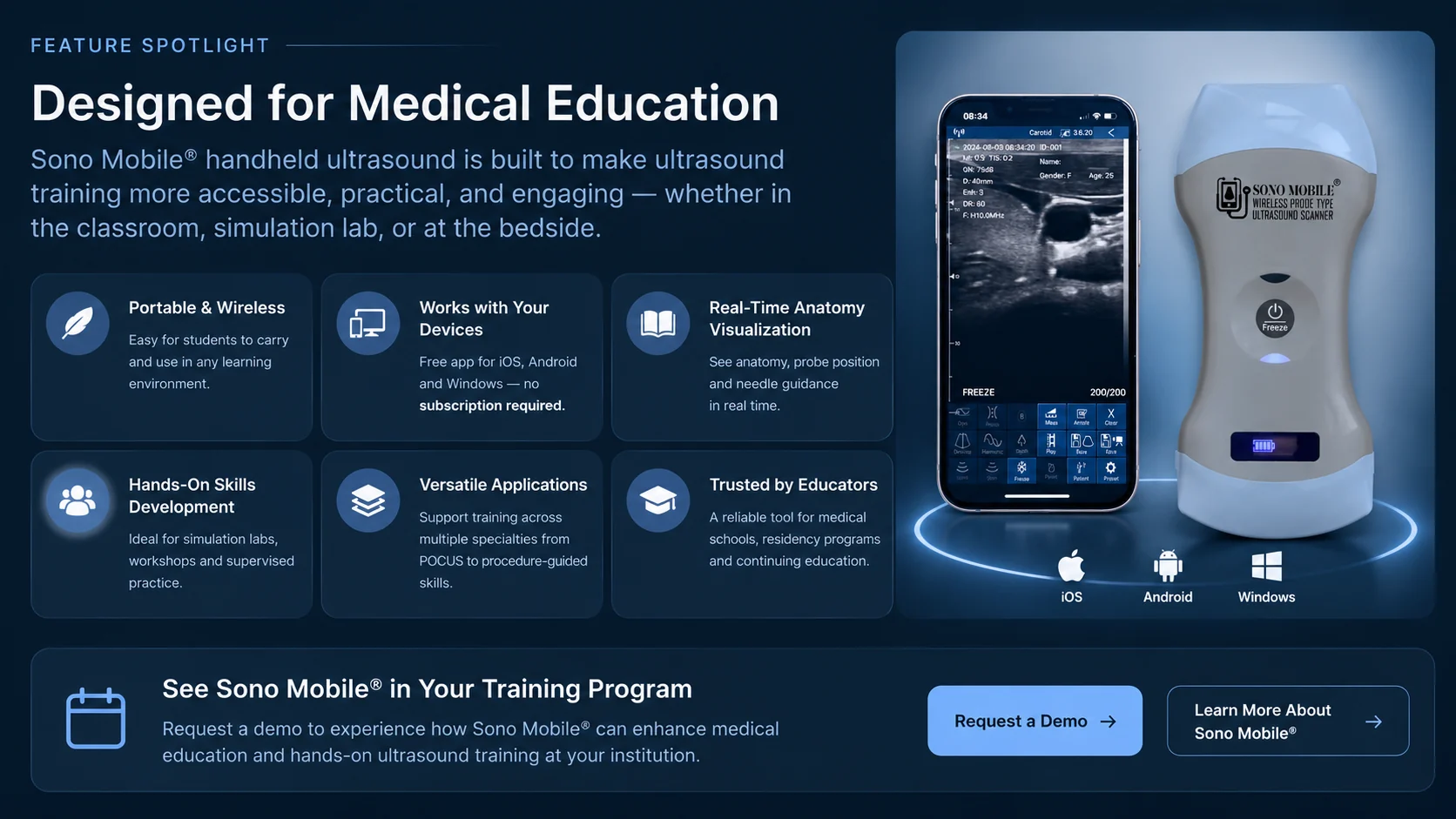
The Sono Mobile® CL64 wireless handheld ultrasound is one example of a portable system that educators may consider for multi-application teaching. Its dual-head configuration combines convex and linear transducers, with phased-array imaging presets, allowing instructors to introduce a range of basic scanning approaches using one device.
Depending on the teaching objective and applicable device specifications, instructors can demonstrate superficial structures with linear imaging, deeper abdominal anatomy with convex imaging, and selected cardiac views using the available phased presets. B-mode, M-mode, Color Doppler, Power Doppler, and Pulsed-Wave Doppler provide additional opportunities to introduce image modes and their limitations.
CL64 can be used with supported mobile and computer platforms through the Sono Mobile software ecosystem. Institutions should verify current platform compatibility, local regulatory status, software functionality, and suitability for their intended educational use before procurement.
Educational positioning: Sono Mobile® CL64 is a teaching tool—not a replacement for qualified instructors, validated curricula, or competency-based assessment. Its suitability for a specific course should be evaluated against that course's learning objectives.
Frequently Asked Questions
Can medical students learn POCUS with a handheld ultrasound device?
Yes. Handheld systems can support introductory POCUS instruction when used within a structured curriculum that includes supervision, practice, feedback, and competency assessment.
Is handheld ultrasound suitable for anatomy education?
It can complement anatomy teaching by showing structures and movement in real time. It should supplement—not replace—other anatomy resources and teaching methods.
What is the difference between ultrasound education and POCUS training?
Ultrasound education can include anatomy, physics, image acquisition, and interpretation across many applications. POCUS training focuses on answering defined bedside clinical questions within the limits of specific protocols.
Which probe types are useful for medical training?
Linear probes are commonly used for superficial structures and vascular access education; convex probes for deeper abdominal structures; and phased-array imaging for selected cardiac applications. The right configuration depends on the curriculum.
Does a handheld device replace formal ultrasound certification?
No. Device ownership or practice does not itself establish clinical competency or professional certification. Training and credentialing requirements depend on the profession, institution, and jurisdiction.
Conclusion: Building Practical Ultrasound Skills
Handheld ultrasound can make medical training more flexible by bringing real-time imaging into teaching sessions, supervised bedside learning, and simulation. The greatest educational value comes when portable equipment is paired with clear learning objectives, qualified instruction, deliberate practice, and meaningful assessment.
For institutions exploring a practical ultrasound teaching setup, the next step is to match the device and training approach to the learners, curriculum, and clinical environment.
Explore Handheld Ultrasound for Your Training Program
Learn about the Sono Mobile® CL64 or contact FAGONEX to discuss an educational demonstration and equipment suitability for your program.
Further Reading and Professional Resources
For curriculum design, clinical scanning guidance, and competency expectations, educators can consult established professional organizations:
- American College of Emergency Physicians (ACEP) — Sonoguide: educational resources on point-of-care ultrasound techniques and applications.
- American Institute of Ultrasound in Medicine (AIUM): professional resources and practice guidance relating to diagnostic medical ultrasound.
- World Federation for Ultrasound in Medicine and Biology (WFUMB): international educational resources and ultrasound guidance.
Educational information only. Clinical scanning, diagnosis, and procedures must follow applicable training, supervision, and professional standards.