

Muscle tear ultrasound provides real-time imaging of skeletal muscle architecture and can help clinicians identify fiber disruption, hematoma, and other changes associated with muscle injury. It also allows assessment of nearby tendons and, when appropriate, observation of tissue movement during a dynamic examination.
Muscle strains and tears occur in sports, physical activity, and everyday clinical practice. Their appearance varies according to injury location, severity, and the time elapsed since injury. This guide explains what a muscle tear can look like on ultrasound, how partial and complete injuries differ, how findings are interpreted, and when MRI or specialist assessment may be needed.
Can Ultrasound Detect a Muscle Tear?
Yes. Ultrasound can detect many muscle tears, particularly in superficial or accessible muscles when the examination is performed by a suitably trained clinician. It can demonstrate alterations in normal muscle architecture, localized fluid collections, and areas of tissue discontinuity.
- Disrupted muscle fibers: Loss of the normal organized muscle pattern.
- Hypoechoic or anechoic regions: Areas that may represent fluid, hemorrhage, or injury-related change.
- Hematoma: A collection of blood within or between muscles, with an appearance that can change over time.
- Retraction or separation: More extensive defects in severe injuries.
- Associated fascial changes: Abnormalities involving the surrounding connective tissue.
Not every muscle injury is visible on ultrasound. Deep injuries, small or early lesions, and complex anatomical regions may be difficult to assess. A negative examination does not always exclude clinically significant injury.
What Does a Muscle Tear Look Like on Ultrasound?
Normal skeletal muscle has a recognizable architecture. On longitudinal views, relatively hypoechoic muscle tissue is traversed by echogenic connective tissue interfaces. Transverse views show a different, more speckled arrangement. A tear can disturb these expected patterns.
Normal Muscle Anatomy on Ultrasound
Understanding normal anatomy is essential before interpreting suspected muscle tears. Clinicians typically scan in both longitudinal and transverse planes and may compare the affected region with the opposite side.
Muscle Tear on Ultrasound: Typical Findings
A suspected tear may appear as focal fiber discontinuity, an irregular hypoechoic region, distortion of the normal fascicular pattern, or a fluid collection. More extensive injuries can produce a visible gap or retracted tissue. Interpretation depends on the specific muscle and clinical circumstances.
Long-Axis and Short-Axis Scanning
Longitudinal scanning follows the direction of muscle fibers and helps demonstrate fiber alignment and disruptions along the muscle. Transverse scanning helps characterize the location and width of an abnormal region. Both views are important because a single scanning plane may miss or misrepresent an injury.
Accurate muscle injury assessment also depends on transducer orientation, controlled probe movements, and image optimization. For step-by-step guidance, explore our musculoskeletal ultrasound scanning techniques guide, covering ultrasound probe positioning, scanning planes, and anisotropy.
Partial vs. Complete Muscle Tears on Ultrasound
Partial Muscle Tear
A partial tear affects a portion of the muscle fibers while other fibers remain intact. Ultrasound may show a localized defect, changes in echogenicity, and surrounding fluid or hemorrhage. Small tears may be subtle, especially early after injury.
Complete Muscle Tear
A complete tear involves full disruption of the affected muscle structure at the injury site. Possible findings include marked discontinuity, separation or retraction of torn ends, and a larger intervening hematoma. The examination should also evaluate nearby musculotendinous structures where clinically relevant.
Muscle Tear Ultrasound Grading
Clinicians use different grading systems to describe muscle injury severity. A simplified three-grade framework is often used for explanation, but it is not a universal ultrasound grading standard; imaging-based classifications may use different definitions.
| Grade | General injury pattern | Possible ultrasound findings |
|---|---|---|
| Grade 1 | Mild injury with limited fiber involvement | Subtle or sometimes absent structural abnormalities |
| Grade 2 | Partial muscle disruption | Focal fiber discontinuity, edema-related change, or hematoma |
| Grade 3 | Severe or complete disruption | Extensive discontinuity, separation, or retraction |
Injury grade should not be assigned from a single image alone. Clinical examination, injury mechanism, imaging findings, and the classification system being used must all be considered.
Dynamic Ultrasound for Muscle and Tendon Injuries
Dynamic musculoskeletal ultrasound allows clinicians to observe structures in motion. Selected movements can help assess tendon excursion, snapping, or positional abnormalities that may not be apparent on static images. In an acute suspected muscle tear, movement should be gentle and guided by clinical judgment to avoid stressing injured tissue.
Related Tendon Injuries: Shoulder and Biceps Ultrasound
Muscle and tendon injuries are related but not interchangeable. A muscle tear involves muscle tissue, whereas a tendon tear involves the connective tissue attaching muscle to bone. The following shoulder examples show how ultrasound can examine nearby tendon structures.
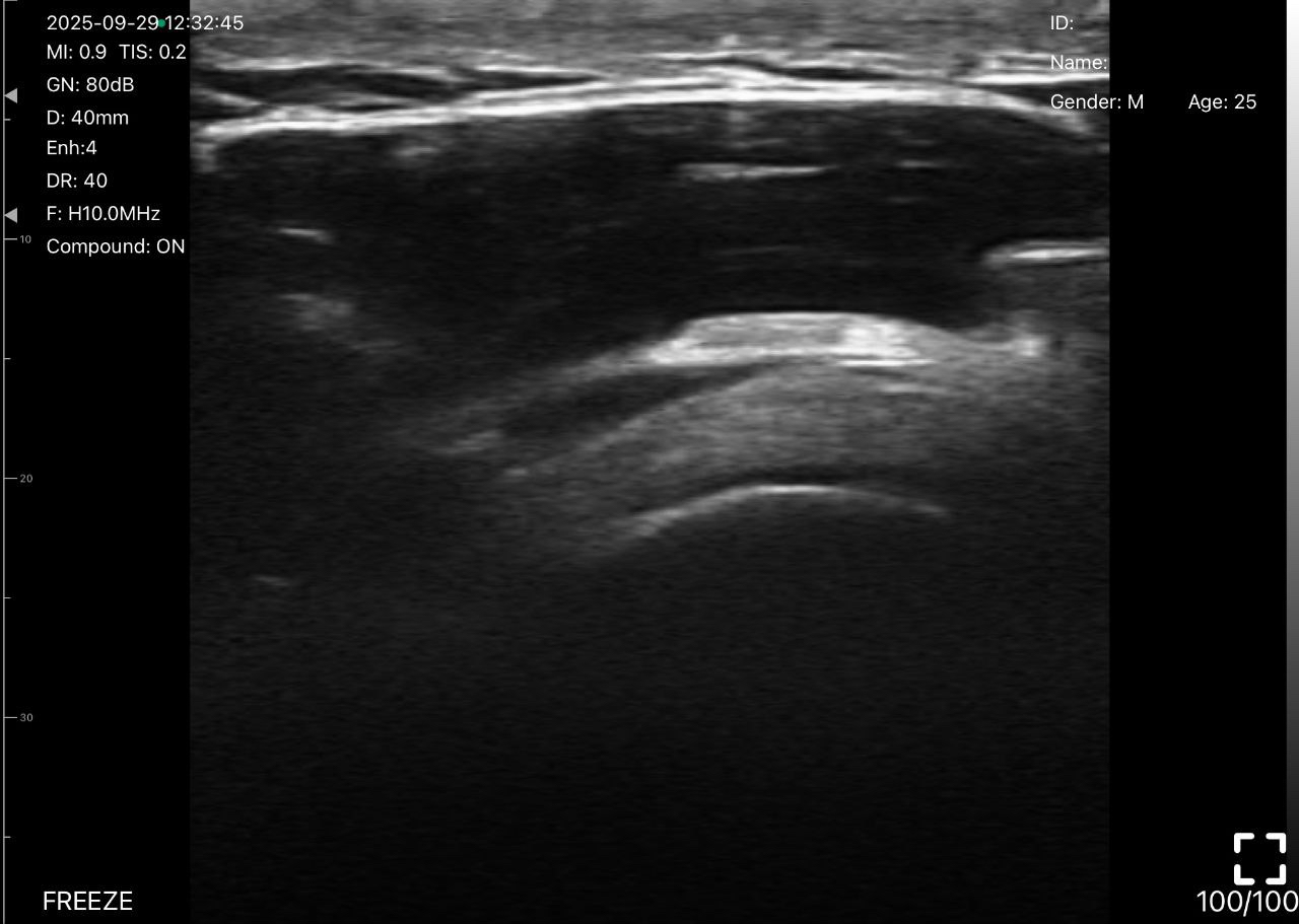
Shoulder Rotator Cuff Tear: Supraspinatus Tendon Ultrasound
The supraspinatus tendon is a major component of the rotator cuff. Ultrasound may demonstrate partial-thickness or full-thickness defects, although accurate interpretation requires appropriate positioning, assessment in multiple planes, and attention to artifacts such as anisotropy. Partial-thickness tears can affect the articular surface, bursal surface, or tendon substance.
Biceps Tendon Ultrasound: Dynamic Assessment of the Long Head
Biceps tendon ultrasound can help examine the long head of the biceps tendon within the bicipital groove. Both short-axis and long-axis views are useful, while carefully selected dynamic shoulder maneuvers may help assess tendon position and suspected instability.
Ultrasound vs. MRI for Muscle Tears
Ultrasound and MRI provide complementary information. The preferred examination depends on the suspected injury, location, clinical question, and available expertise.
| Ultrasound | MRI |
|---|---|
| Real-time and dynamic imaging | Detailed cross-sectional imaging |
| Portable and often available at the point of care | Broad field of view, including deep structures |
| Useful for accessible superficial muscles and tendons | Often helpful for complex, deep, or inconclusive injuries |
| Results depend strongly on operator skill and scanning technique | Less dependent on real-time probe positioning |
MRI may be appropriate when ultrasound findings are inconclusive, when a deep injury is suspected, or when a more comprehensive anatomical assessment is required.
Handheld Ultrasound for Muscle and Tendon Assessment
Portable systems can support musculoskeletal examinations in appropriate clinical settings. High-frequency linear imaging is commonly used for accessible superficial muscles and tendons, while the optimal probe and settings depend on the depth and anatomy being assessed.
The Sono Mobile® CL64 offers a wireless handheld format with linear imaging capability. Explore the Sono Mobile® CL64 for multi-application scanning and the Sono Mobile® L4 linear ultrasound for superficial structures. You can also compare devices in the handheld ultrasound product category. The videos above illustrate selected muscle and tendon scanning applications using Sono Mobile® equipment. Image interpretation remains dependent on clinical training and examination technique.
Explore Musculoskeletal Ultrasound with Sono Mobile®
Explore clinical applications, compare wireless handheld ultrasound devices, or request a product demonstration for your practice.
Request a Demo →Explore Devices →Learn more about MSK ultrasound and point-of-care ultrasound.
Frequently Asked Questions About Muscle Tear Ultrasound
Will ultrasound show a muscle tear?
Ultrasound can show many muscle tears by demonstrating changes in fiber continuity, tissue architecture, and associated fluid collections. Detection depends on injury size, depth, timing, and operator expertise.
Can an ultrasound detect a partial muscle tear?
Yes. Partial tears may appear as localized fiber disruption or abnormal echotexture, although subtle injuries can be difficult to identify.
What does a torn muscle look like on ultrasound?
Possible findings include interrupted muscle fibers, focal hypoechoic regions, hematoma, and—in severe cases—separation or retraction of tissue.
Is ultrasound or MRI better for muscle tears?
Neither is universally better. Ultrasound provides dynamic, real-time assessment of accessible structures; MRI can be valuable for deep, complex, or uncertain injuries.
Can Doppler ultrasound diagnose a muscle tear?
Doppler may provide supplementary information about vascularity in selected cases, but it cannot establish the presence or grade of a tear by itself.
Are a biceps tendon tear and a biceps muscle tear the same?
No. A tendon tear involves the fibrous tendon, while a muscle tear involves muscle tissue. Their anatomy, imaging appearances, and clinical implications differ.
Conclusion
Muscle tear ultrasound is a useful tool for evaluating muscle fiber architecture, localized defects, and associated soft-tissue abnormalities. Its real-time capabilities also support selected dynamic assessments of nearby tendons. Accurate interpretation requires appropriate technique, clinical correlation, and recognition of the modality’s limitations.
For additional applications and scanning considerations, explore FAGONEX’s MSK ultrasound specialty page, the POCUS clinical guide, and the broader medical specialties overview.
Clinical References and Further Reading
- Radiopaedia — Muscle Tear. Reference overview of imaging findings in muscle injury.
- Radiopaedia — Muscle Injury. Overview of muscle injury imaging and terminology.
- American Institute of Ultrasound in Medicine (AIUM) — Musculoskeletal Ultrasound Resources. Professional guidance and educational resources; check current availability of the linked page.
Medical disclaimer: This content is intended for healthcare education only and does not replace professional clinical assessment, formal imaging interpretation, or individualized treatment decisions. Videos are labeled according to their supplied descriptions and should not be treated as independent confirmation of a diagnosis.