
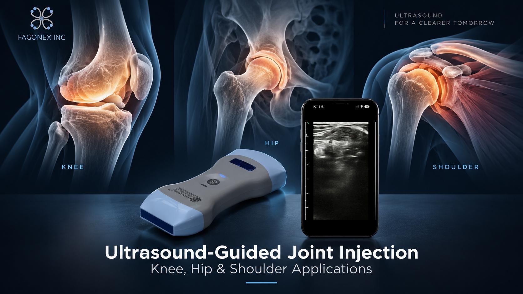
An ultrasound guided joint injection uses real-time imaging to help trained clinicians identify an intra-articular target, assess surrounding anatomy, and monitor needle positioning during selected joint procedures. This clinical guide focuses on knee, hip, and shoulder injections, the evidence for placement accuracy, and the limitations and safety considerations that influence clinical decision-making.
For a broader overview of joint and soft-tissue procedures, see our ultrasound-guided injections guide . This article concentrates specifically on intra-articular joint applications.
What Is an Ultrasound-Guided Joint Injection?
An intra-articular injection delivers a selected medication or other clinically indicated substance into a joint space. In an ultrasound-guided procedure, the clinician uses sonographic imaging to evaluate the target joint and adjacent structures before and, when appropriate, during needle advancement.
Depending on the clinical indication, joint injections may be considered in the assessment or management of certain inflammatory or degenerative joint conditions. The choice of treatment must follow a clinical diagnosis, an evaluation of potential benefits and harms, and shared decision-making.
Ultrasound is also used for joint aspiration, where synovial fluid is collected for diagnostic or therapeutic purposes. Although aspiration and injection may involve related imaging considerations, they are distinct clinical interventions.
Clinicians interested in broader anatomical assessment can explore the FAGONEX musculoskeletal ultrasound specialty and our guide to musculoskeletal ultrasound fundamentals .
Why Use Ultrasound Guidance for Joint Injections?
Ultrasound provides real-time visualization of accessible joint anatomy, adjacent soft tissues, and selected aspects of the needle pathway. It may be particularly useful when surface landmarks are difficult to identify or when the joint lies deep beneath surrounding tissue.
Target Identification
Sonographic imaging can help distinguish the intended joint space from nearby muscles, tendons, vessels, and other tissues.
Dynamic Imaging
Clinicians can observe anatomy in real time and reassess the relationship between the planned target and surrounding structures.
Needle Visualization
Appropriate ultrasound techniques may support observation of the needle relative to the target, although continuous visualization is not always achievable.
Individualized Assessment
Imaging may assist planning in patients with challenging anatomy, altered landmarks, or selected joint abnormalities.
A 2024 umbrella review reported that ultrasound-guided musculoskeletal injections were generally more accurate than landmark-guided procedures across several anatomical sites. However, improvements in pain relief and functional outcomes varied between conditions and studies. [1]
Ultrasound-Guided Knee Injection
An ultrasound guided joint injection of the knee is a commonly studied application of image-guided intra-articular procedures. Ultrasound can demonstrate selected joint recesses, fluid collections, and adjacent soft-tissue anatomy. This can help clinicians identify a suitable target when conventional landmarks are difficult to assess.
Clinical applications may include selected interventions for symptomatic knee osteoarthritis and other conditions where intra-articular treatment is appropriate. The medication or intervention selected should be based on current evidence, clinical assessment, and professional guidance.
What Does the Evidence Show?
A review examining intra-articular knee injections found higher placement accuracy with ultrasound guidance than with anatomical landmark guidance. The reported knee-specific comparison was approximately 95.8% versus 77.8% across the reviewed studies. These are pooled research findings, not guaranteed accuracy rates for individual clinicians or procedures. [2]
A separate meta-analysis of randomized trials involving knee osteoarthritis reported advantages for ultrasound guidance in selected procedural pain, follow-up pain, and functional outcomes. The number of included trials was limited, and the findings should not be generalized to every joint condition or medication. [3]
For related sports-related knee and musculoskeletal assessments, visit our sports medicine ultrasound guide .
Ultrasound-Guided Hip Injection
The hip is a relatively deep joint surrounded by substantial muscle and soft-tissue structures. Its depth and proximity to important neurovascular anatomy can make surface-landmark techniques more challenging than those used for certain superficial joints.
For an ultrasound guided joint injection of the hip, real-time imaging allows a trained clinician to identify relevant anatomical landmarks and assess the intended intra-articular target. Adequate imaging penetration, probe selection, and operator experience are particularly important for deeper structures.
Accuracy of Hip Joint Injections
A systematic review and meta-analysis reported weighted mean placement accuracy of 100% for ultrasound-guided hip injections and 72% for landmark-guided injections in the included studies. These findings reflect specific research samples and should not be interpreted as universal clinical success rates. [4]
Ultrasound may not be the optimal imaging modality in every case. Deep anatomy, limited acoustic windows, previous surgery, and patient-specific factors can influence whether ultrasound or an alternative guidance method is appropriate.
Ultrasound-Guided Shoulder Injection
Shoulder pain can arise from several anatomical structures. An ultrasound guided joint injection targeting the shoulder must therefore be understood in relation to the specific intended target, rather than as one uniform procedure.
Glenohumeral Joint Injection
The glenohumeral joint is the principal ball-and-socket articulation of the shoulder. Ultrasound may help identify relevant joint anatomy and guide selected intra-articular interventions.
A systematic review published in 2025 found that ultrasound-guided glenohumeral injections generally achieved greater placement accuracy than landmark-guided injections, but available comparative clinical studies did not consistently demonstrate superior pain or functional outcomes. [5]
Acromioclavicular (AC) Joint Injection
The acromioclavicular joint is a smaller, more superficial articulation between the clavicle and acromion. Its anatomical characteristics differ from those of the glenohumeral joint, and its clinical indications should be evaluated separately.
Although ultrasound can help visualize the AC joint, the available evidence does not establish that ultrasound guidance improves patient-centered outcomes for every AC joint injection.
Joint Versus Periarticular Shoulder Injections
A subacromial-subdeltoid bursa injection or biceps tendon-sheath injection is not the same as a glenohumeral intra-articular injection. Distinguishing these targets is essential for accurate clinical communication and interpretation of research.
A Cochrane review of glucocorticoid injections for shoulder pain found little or no clinically important improvement in pain or function with image guidance compared with injections performed without imaging. The review included several shoulder conditions and injection targets, so its findings should be interpreted within that context. [6]
Ultrasound-Guided vs. Landmark-Guided Joint Injections
Both ultrasound-guided and landmark-guided approaches are used in clinical practice. The preferred method depends on the intended target, patient anatomy, clinician expertise, available equipment, and evidence relevant to the intervention.
| Joint | Imaging considerations | Clinical context |
|---|---|---|
| Knee | Ultrasound can visualize selected recesses, effusions, and soft tissues. | Evidence supports improved placement accuracy; symptomatic benefits vary. |
| Hip | Deep anatomy requires sufficient penetration and appropriate imaging. | Accuracy evidence favors ultrasound over landmark guidance in reviewed studies. |
| Glenohumeral | Imaging helps identify the intended shoulder joint target. | Greater accuracy does not consistently translate into superior clinical outcomes. |
| AC Joint | A small superficial joint with distinct anatomical considerations. | Evidence of superior patient-centered outcomes remains limited. |
Fluoroscopy and other imaging modalities may also be considered for selected procedures. Ultrasound has the advantage of real-time soft-tissue visualization without ionizing radiation, but it does not replace every alternative technique.
Does Better Injection Accuracy Mean Better Pain Relief?
Not necessarily. Placement accuracy describes whether the intended anatomical target is reached. Clinical effectiveness involves different outcomes, including pain reduction, physical function, patient satisfaction, treatment duration, and adverse events.
These outcomes are influenced by the underlying diagnosis, injected substance, disease severity, rehabilitation, concurrent treatments, and other patient-specific factors.
Some studies show clinical benefits from ultrasound guidance, while others report minimal differences compared with landmark-guided interventions. A balanced interpretation must distinguish technical success from meaningful improvement in symptoms.
Safety, Contraindications and Clinical Limitations
Ultrasound guidance does not make joint injection risk-free. Potential complications vary with the procedure and may include infection, bleeding, injury to nearby structures, medication-related adverse effects, or failure to achieve the intended clinical outcome.
Before any intervention, the responsible clinician should assess the indication, relevant medical history, allergies, anticoagulant use, suspected infection, and other patient-specific risk factors. Informed consent, appropriate aseptic precautions, and local clinical protocols remain essential.
Limitations of Ultrasound Imaging
- Image quality may decrease with increasing tissue depth or unfavorable acoustic windows.
- Bone and other structures can create acoustic shadowing.
- Needle visibility depends on multiple technical and anatomical factors.
- Ultrasound interpretation is operator-dependent.
- Some clinical situations require alternative imaging or specialist care.
For a broader discussion of image-guided interventions, including periarticular procedures, visit our ultrasound-guided injections overview .
Handheld Ultrasound for Joint Injection Assessment and Planning
Ultrasound system selection should reflect the anatomical target, depth, required image quality, and clinical workflow. High-frequency linear transducers are commonly useful for accessible superficial joints and soft tissues, while deeper structures may require lower-frequency imaging and sufficient penetration.
A suitable system should provide reliable anatomical visualization, appropriate image optimization, practical ergonomics, and a workflow compatible with the clinical environment. Doppler capabilities may assist assessment of nearby vessels when clinically indicated.
Portable and handheld ultrasound systems can support selected musculoskeletal and point-of-care workflows when their capabilities match the intended application. They should not be assumed equivalent for every intervention or imaging depth.
Explore Handheld Ultrasound for Musculoskeletal Care
Sono Mobile® CL64 combines linear and convex transducers in a wireless handheld platform, supporting a range of superficial and deeper ultrasound assessments. Suitability for a particular procedure depends on the imaging requirements, clinical indication, and operator training.
Explore the broader role of ultrasound in pain management or review device capabilities before selecting equipment for your practice.
How Joint Injections Fit Into Musculoskeletal Pain Management
An ultrasound guided joint injection may be one component of a broader musculoskeletal pain management plan. Depending on the diagnosis, care may involve exercise therapy, rehabilitation, medication, patient education, activity modification, or specialist evaluation.
Ultrasound may also support assessment of joint effusion, synovial abnormalities, and surrounding soft tissues when appropriate. Imaging findings should always be interpreted alongside the history, physical examination, and other clinical information.
Explore related applications through our orthopedic ultrasound guide , physiotherapy ultrasound resources , and point-of-care ultrasound (POCUS) pillar page.
Frequently Asked Questions
What is an ultrasound guided joint injection?
It is an intra-articular procedure in which a trained clinician uses ultrasound imaging to assess joint anatomy and support accurate placement of a needle toward the intended target.
Are ultrasound-guided knee injections more accurate?
Systematic reviews generally report higher placement accuracy with ultrasound guidance than with landmark-based techniques for knee joint injections. Individual results depend on the procedure and operator.
Why is ultrasound used for hip injections?
The hip is a deep joint. Ultrasound can help visualize relevant anatomy and support target localization. Evidence from comparative studies suggests higher placement accuracy than landmark guidance.
Is a glenohumeral injection the same as a shoulder bursa injection?
No. A glenohumeral injection targets the shoulder joint cavity, while a bursa injection targets a separate periarticular structure.
Does ultrasound guidance guarantee better pain relief?
No. Greater anatomical accuracy does not necessarily lead to better pain relief or long-term function. Clinical outcomes vary by condition, treatment, and patient.
Can handheld ultrasound be used for joint injections?
Selected handheld systems may support relevant anatomical visualization when image quality, transducer configuration, and clinical requirements are suitable. Operator training and institutional standards remain essential.
Conclusion
Ultrasound guided joint injection provides real-time anatomical information that can support selected intra-articular procedures involving the knee, hip, shoulder, and other joints. Evidence generally favors ultrasound for placement accuracy, although improvements in symptoms and long-term outcomes are not consistent across all indications.
The most appropriate approach depends on the clinical diagnosis, joint anatomy, available evidence, patient preferences, and the experience of the treating clinician.
Scientific References
- Comparison of ultrasound- vs. landmark-guided injections for musculoskeletal pain: an umbrella review. Journal of Rehabilitation Medicine, 2024. PubMed .
- Clinical utility of ultrasound guidance for intra-articular knee injections: a review. PubMed .
- Comparison of ultrasound guidance with landmark guidance for symptomatic benefits in knee, hip and hand osteoarthritis: systematic review and meta-analysis. Full Text .
- Ultrasound-guided hip joint injections are more accurate than landmark-guided injections: a systematic review and meta-analysis. British Journal of Sports Medicine. PubMed .
- Accuracy and Efficacy of Ultrasound-Guided Glenohumeral Joint Injections: A Systematic Review. PubMed .
- Zadro J, et al. Image-guided glucocorticoid injection versus injection without image guidance for shoulder pain. Cochrane Database of Systematic Reviews, 2021. Cochrane Review .
Medical editorial note: This article is intended for professional education only. It does not provide a procedural injection protocol or replace supervised clinical training, professional guidelines, informed consent, or individualized medical judgment. All invasive procedures should be performed by appropriately qualified healthcare professionals.