Medical imaging plays a crucial role in diagnosing and managing a wide range of conditions. Among the many modalities available, ultrasound and magnetic resonance imaging (MRI) stand out for their versatility and widespread use. While both are indispensable in modern healthcare, they differ significantly in technology, applications, and patient experience. This article explores the key differences between ultrasound and MRI, providing insights into their capabilities and helping clinicians and patients make informed decisions.
Introduction to Ultrasound and MRI
Ultrasound and MRI are diagnostic imaging techniques used to visualize internal structures of the body. Ultrasound employs high-frequency sound waves to generate images of soft tissues, blood vessels, and organs. It is widely known for its use in obstetrics, such as finding out a baby’s sex with an ultrasound, but its applications extend far beyond that, including musculoskeletal and cardiovascular imaging.
MRI, on the other hand, uses powerful magnetic fields and radio waves to create detailed images of soft tissues, bones, and organs. Unlike ultrasound, MRI provides a three-dimensional view of structures and is often used for complex diagnostic evaluations.
Both modalities are valuable tools, but their differences make each suited for specific clinical situations.
How Ultrasound Works
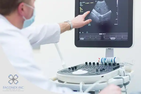
Ultrasound imaging relies on sound waves to produce real-time images of the body. A handheld device called a transducer emits high-frequency sound waves that travel through the body and bounce off tissues, creating echoes. These echoes are captured by the transducer and converted into visual images on a monitor.
Advantages of Ultrasound
- Real-Time Imaging: Ideal for observing dynamic processes, such as blood flow or joint movement.
- Portability: Devices like portable ultrasound machines and handheld ultrasounds, including those by Sono Mobile™️, make it easy to perform imaging at the bedside or in remote locations.
- Safety: Ultrasound does not involve ionizing radiation, making it safe for repeated use, especially in sensitive populations like pregnant women.
- Cost-Effective: Ultrasound is generally more affordable than other imaging modalities, increasing its accessibility.
How MRI Works
Magnetic resonance imaging (MRI) utilizes a combination of a strong magnetic field and radiofrequency pulses to generate detailed cross-sectional images of the body. The technology excites hydrogen atoms in the body, causing them to emit signals. These signals are captured by the MRI machine and processed to create high-resolution images.
Advantages of MRI
- Exceptional Detail: MRI excels at imaging soft tissues, such as the brain, spinal cord, and joints.
- No Radiation Exposure: Like ultrasound, MRI does not use ionizing radiation, making it a safer option for long-term monitoring.
- 3D Imaging Capabilities: MRI provides comprehensive three-dimensional views, which are especially valuable for surgical planning.
- Versatility: From neurological disorders to orthopedic injuries, MRI offers unparalleled diagnostic precision.
Comparison of Imaging Capabilities
Both ultrasound and MRI are used to evaluate soft tissues and organs, but their strengths vary significantly.
Soft Tissue Imaging
- Ultrasound: Excellent for visualizing superficial tissues such as tendons, ligaments, and muscles. Ideal for dynamic assessments, such as tracking joint movement or blood flow in real time.
- MRI: Superior for deep and complex structures, including the brain, spinal cord, and cartilage. Provides unparalleled detail in conditions like multiple sclerosis or ligament tears.
Bone Imaging
- Ultrasound: Limited in bone imaging, primarily detecting surface irregularities or guiding injections near bones.
- MRI: Offers comprehensive views of bone marrow and adjacent soft tissues, aiding in diagnosing fractures, tumors, and infections.
Dynamic vs. Static Imaging
- Ultrasound: Real-time imaging makes it ideal for interventional procedures and functional assessments.
- MRI: Produces static images, but the detail is unmatched for diagnosing chronic or complex conditions.
Patient Experience
The patient experience varies significantly between ultrasound and MRI due to differences in procedure length, comfort, and setting.
Ultrasound Experience
Ultrasound is typically quick and non-invasive. Patients lie on an examination table while a technician applies a gel to the skin and moves the transducer over the area being examined. The process is painless, and results are often available immediately.
- Duration: Most exams take 15–30 minutes.
- Environment: Performed in a variety of settings, from clinics to emergency rooms, thanks to devices like point-of-care ultrasound (POCUS) systems.
- Comfort: Generally comfortable and accessible for all age groups.
MRI Experience
MRI requires the patient to lie still inside a large, enclosed scanner. The procedure is noisier and may last longer, which can cause discomfort for some individuals, especially those with claustrophobia.
- Duration: Exams can take 30–90 minutes.
- Environment: Requires specialized facilities with trained personnel.
- Comfort: May be challenging for patients with metallic implants, anxiety, or sensitivity to loud noises.
Safety Considerations
Both ultrasound and MRI are considered safe, but certain factors should be considered.
Ultrasound Safety
- No Radiation: Ultrasound uses sound waves, posing no risk of ionizing radiation.
- Repetitive Use: Safe for frequent imaging, even during pregnancy.
- Limitations: High-frequency sound waves may not penetrate deep tissues effectively, limiting diagnostic capabilities in obese patients.
- MRI Safety
- No Radiation: MRI also avoids ionizing radiation, making it suitable for repeated imaging.
- Metal Implants: Patients with pacemakers, metal prosthetics, or other implants may not be eligible due to the strong magnetic field.
- Contrast Agents: Some MRI scans require the use of gadolinium-based contrast agents, which carry minimal risk of allergic reactions or kidney complications.
Cost and Accessibility
Cost and accessibility are important considerations for both patients and healthcare providers.
Ultrasound
- Affordability: Ultrasound is one of the most cost-effective imaging modalities, with lower operational and maintenance expenses.
- Portability: Devices like handheld ultrasound machines enable imaging in remote or underserved areas.
- Availability: Widely available in outpatient clinics, hospitals, and even private practices.
- MRI
- High Cost: MRI machines are expensive to purchase and maintain, resulting in higher costs for patients and providers.
- Limited Accessibility: MRI facilities are less common in rural areas, requiring patients to travel to specialized centers.
- Waiting Times: Due to limited availability, patients may face longer wait times for non-urgent MRI scans.
Clinical Applications
Ultrasound Applications
Ultrasound is commonly used for:
Obstetrics and Gynecology: Monitoring pregnancy, assessing fetal development, and finding out a baby’s sex with an ultrasound.
- Musculoskeletal Conditions: Diagnosing sprains, tendon tears, and guiding injections.
- Emergency Medicine: Evaluating internal bleeding, fractures, and organ damage at the point of care.
- Cardiology: Assessing heart function and blood flow with echocardiography.
- MRI Applications
- MRI is preferred for:
- Neurology: Diagnosing brain and spinal cord conditions, such as tumors or multiple sclerosis.
- Orthopedics: Detecting ligament tears, cartilage damage, and bone infections.
- Oncology: Imaging soft tissue tumors and tracking cancer progression.
- Cardiovascular Imaging: Evaluating heart structure, blood vessels, and congenital abnormalities.
Technological Advances
Both ultrasound and MRI are benefiting from technological innovations, improving their diagnostic and therapeutic capabilities.
Ultrasound Advances
- Handheld and Portable Devices: Compact systems like those from Sono Mobile™️ allow imaging in diverse settings, from ambulances to sports fields.
- AI Integration: Artificial intelligence enhances image interpretation, reducing variability between operators.
- 3D and 4D Imaging: Advancements provide more detailed anatomical views, improving diagnostic accuracy.
- MRI Advances
- High-Field Scanners: Newer machines with stronger magnets produce sharper images in less time.
- Functional MRI (fMRI): Used for mapping brain activity and planning surgeries.
- Hybrid Systems: Combining MRI with PET or CT scans provides comprehensive diagnostic information.
Conclusion: Which Modality to Choose?
The choice between ultrasound and MRI depends on the clinical situation, patient needs, and resource availability.
Choose Ultrasound When:
- Real-time imaging is required for dynamic assessments or interventional procedures.
- Cost, portability, and accessibility are key considerations.
- Diagnosing conditions like soft tissue injuries, pregnancy-related issues, or organ evaluations.
- Choose MRI When:
- Detailed visualization of complex structures like the brain, spine, or joints is needed.
- Long-term monitoring of chronic conditions or cancer is required.
- High diagnostic precision outweighs cost and time constraints.
Ultimately, both modalities have unique strengths, making them complementary rather than competitive. By leveraging the best of both technologies, clinicians can ensure accurate diagnoses and optimal patient care.
Explore advanced ultrasound solutions at FAGONEX Cooperation Inc., where we specialize in point-of-care ultrasound (POCUS) devices designed to meet modern healthcare demands.
One Response
I have been surfing online more than 3 hours lately, yet I
by no means found any interesting article like yours. It’s beautiful
price enough for me. In my view, if all website owners and bloggers made
good content material as you probably did, the internet will likely be
a lot more helpful than ever before.