
What Is the RUSH Exam?
The RUSH exam — Rapid Ultrasound for Shock and Hypotension — is a structured point-of-care ultrasound approach used to rapidly evaluate patients with undifferentiated hypotension or shock. It combines focused assessment of the heart, intravascular volume clues, lungs, abdomen, aorta and selected venous structures to help clinicians narrow the differential diagnosis while resuscitation is underway.
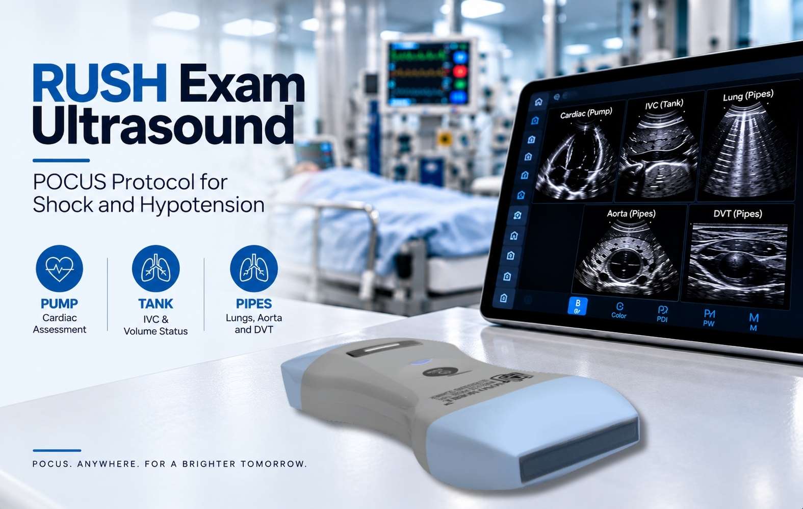
A widely taught framework organizes the RUSH exam ultrasound into three physiologic questions: assess the Pump, evaluate the Tank, and inspect the Pipes.
Why Use the RUSH Exam in POCUS?
Shock can have several mechanisms, including hypovolemic, cardiogenic, distributive and obstructive causes. These categories can overlap, and the initial presentation may not immediately reveal the dominant mechanism.
The RUSH exam POCUS protocol provides a repeatable way to collect focused ultrasound information during assessment of a hypotensive patient. Instead of treating one ultrasound sign as a diagnosis, findings are integrated with the history, physical examination, vital signs, laboratory data and response to treatment.
Point-of-care ultrasound is particularly suited to this setting because imaging can be performed at the bedside and repeated as the patient's condition changes. Learn more about point-of-care ultrasound (POCUS).
HI-MAP and Pump–Tank–Pipes
The original RUSH approach is often remembered using the HI-MAP mnemonic: Heart, IVC, Morison's pouch/FAST, Aorta and Pneumothorax. The protocol later evolved into the broader Pump–Tank–Pipes framework, connecting focused ultrasound findings with cardiovascular physiology.
Pump
Evaluate cardiac activity and gross ventricular function, look for pericardial fluid and assess findings that may suggest right-heart strain in the appropriate clinical setting.
Tank
Assess intravascular volume clues and potential fluid loss or accumulation using IVC, lung, pleural and abdominal views.
Pipes
Examine major vascular structures when indicated, particularly the abdominal aorta and lower-extremity veins.
RUSH Exam Ultrasound Views at a Glance
| Component | Common Views | Focused Question |
|---|---|---|
| Pump | Parasternal, apical or subcostal cardiac views | What does global cardiac function and chamber appearance suggest? |
| Tank | IVC | What filling or volume-status clues are present when interpreted in context? |
| Tank | Anterior and lateral lung | Are B-lines, pleural fluid or signs associated with pneumothorax present? |
| Tank | RUQ / Morison's pouch and other FAST windows | Is free intraperitoneal fluid visible? |
| Pipes | Abdominal aorta | Is significant aortic pathology suspected or visualized? |
| Pipes | Lower-extremity venous compression | Is proximal DVT present when the clinical scenario warrants assessment? |
Cardiac Assessment in the RUSH Exam
The cardiac component asks whether the heart provides clues to the patient's hemodynamic state. Focused cardiac POCUS may include subcostal, parasternal and apical windows depending on the patient, operator skill and urgency.
The examination commonly considers gross left-ventricular systolic function, relative right- and left-ventricular size, pericardial fluid and the overall pattern of cardiac filling. These observations must be interpreted together.
IVC Assessment and Volume-Status Clues
Inferior vena cava imaging is frequently incorporated into the RUSH protocol. The IVC can provide information related to right-sided filling conditions, but its size and respiratory behavior should not be interpreted as a stand-alone measurement of circulating volume.
Ventilation mode, respiratory effort, right-heart function and other physiologic factors can change IVC appearance. The IVC is therefore most useful when integrated with cardiac and lung findings and the overall clinical picture.
Lung Ultrasound: B-Lines, Pleural Fluid and Pneumothorax Clues
Lung ultrasound extends the RUSH exam beyond the circulation itself. A focused lung assessment can identify patterns that help refine the differential diagnosis of shock and respiratory distress.
B-Lines
Multiple B-lines in the appropriate distribution can indicate an interstitial syndrome. In a hypotensive patient, this finding becomes more useful when interpreted alongside cardiac function, IVC appearance and the broader clinical picture.
Pleural Effusion
Pleural fluid can also be detected at the bedside. Its presence, distribution and associated findings may contribute additional information when evaluating circulatory or respiratory compromise.
Pneumothorax
In the appropriate clinical setting, lung POCUS can assess findings associated with pneumothorax, including lung sliding and other pleural signs. These findings should be interpreted according to operator training and the clinical context.
Morison's Pouch and Abdominal Free Fluid
The abdominal component of the RUSH exam overlaps with FAST principles. The right upper quadrant, including the hepatorenal recess or Morison's pouch, is an important window for detecting free intraperitoneal fluid.
In a patient with hypotension, free fluid may substantially change the working differential diagnosis, although its meaning depends on the clinical setting. Additional FAST windows can be examined when indicated.
Aorta and DVT Assessment in the RUSH Protocol
The “Pipes” component focuses on major vascular structures. Depending on the patient's presentation, this may include assessment of the abdominal aorta and compression ultrasound of proximal lower-extremity veins.
Abdominal Aorta
Focused aortic ultrasound can be particularly relevant when hypotension is accompanied by abdominal, flank or back symptoms or when aortic pathology is otherwise part of the differential diagnosis.
Deep Venous Thrombosis
Lower-extremity venous compression may be added when thromboembolic disease is clinically suspected. A positive DVT study can add important information, but the examination must still be interpreted within the patient's presentation.
How RUSH Findings May Differ Across Shock States
The goal of the RUSH exam is not to diagnose shock from a single image. Its value comes from combining several ultrasound observations into a physiologic pattern. Findings can vary substantially, particularly in mixed shock.
Hypovolemic
Findings may suggest reduced filling, while abdominal or other views may reveal a potential source of fluid loss depending on the clinical scenario.
Cardiogenic
Cardiac dysfunction and lung findings compatible with pulmonary congestion may contribute to the overall pattern.
Obstructive
Focused ultrasound may reveal findings associated with conditions such as tamponade or tension pneumothorax, or findings that increase concern for pulmonary embolism in the appropriate context.
Distributive
Ultrasound findings may be less specific and can evolve during resuscitation. Serial assessment and integration with clinical data are especially important.
RUSH vs FAST vs eFAST: What Is the Difference?
| Protocol | Primary Focus | Typical Context |
|---|---|---|
| RUSH | Multiorgan assessment of undifferentiated shock/hypotension | Hemodynamic instability |
| FAST | Focused search for free fluid in standard trauma windows | Trauma assessment |
| eFAST | FAST plus thoracic assessment for pneumothorax/hemothorax | Expanded trauma assessment |
FAST views can form part of a RUSH examination, but the protocols are not interchangeable. RUSH has a broader hemodynamic objective and combines cardiac, pulmonary, abdominal and vascular information.
What Does the Evidence Say About the RUSH Exam?
Published studies and systematic reviews support the usefulness of multiorgan POCUS in patients with undifferentiated shock. A systematic review and meta-analysis reported favorable overall diagnostic performance for RUSH in differentiating shock states.
Other evidence indicates that RUSH may perform better for ruling in certain causes of shock than for definitively excluding individual etiologies. A reassuring or incomplete RUSH examination should therefore not prevent further investigation when clinical concern remains.
Why Serial POCUS Matters in Shock
Shock is dynamic. Cardiac filling, lung findings and other sonographic signs can change after fluids, vasoactive therapy, ventilation or other interventions. This makes repeat bedside ultrasound useful when clinicians need to reassess physiology over time.
The most useful sequence is not necessarily identical for every patient. Clinicians may prioritize the highest-yield views first and expand the examination according to the presentation, available time and initial findings.
Where Is RUSH Exam POCUS Used?
RUSH is most closely associated with acute and bedside care environments where rapid assessment of hemodynamic instability is required.
RUSH at the Bedside with Handheld Ultrasound
A RUSH workflow may require rapid movement between cardiac, abdominal, vascular and lung windows. Handheld ultrasound can support this bedside workflow by keeping imaging accessible at the point of care.
Sono Mobile® CL64 combines convex, linear and phased-array scanning capabilities in a wireless handheld platform, supporting multiple ultrasound applications from a single device.
Explore Sono Mobile® CL64RUSH Exam Ultrasound FAQ
What does RUSH stand for in ultrasound?
RUSH stands for Rapid Ultrasound for Shock and Hypotension. It is a structured POCUS approach used during evaluation of patients with undifferentiated hypotension or shock.
What are the components of the RUSH exam?
The expanded RUSH framework is commonly organized as Pump, Tank and Pipes. It includes focused cardiac assessment, IVC and lung evaluation, assessment for free abdominal fluid and selected vascular examinations such as the abdominal aorta and lower-extremity veins when indicated.
What is HI-MAP in the RUSH exam?
HI-MAP is a mnemonic for Heart, IVC, Morison's pouch/FAST, Aorta and Pneumothorax. It represents an earlier framework for organizing the RUSH examination.
Is the RUSH exam the same as FAST?
No. FAST primarily evaluates for free fluid in trauma. RUSH incorporates cardiac, pulmonary, abdominal and vascular POCUS to assess undifferentiated shock and hypotension. FAST views may be included within a RUSH examination.
Can RUSH determine the cause of shock by itself?
No. RUSH findings are interpreted with the patient's history, examination, vital signs, laboratory data and other diagnostic testing. It is a focused bedside assessment rather than a replacement for comprehensive diagnostic evaluation.
Can handheld ultrasound be used for a RUSH exam?
A handheld ultrasound system can support RUSH when it provides the imaging capabilities required for relevant cardiac, lung, abdominal and vascular views and is used by an appropriately trained clinician.
References and Further Reading
- American College of Emergency Physicians (ACEP). Rapid Ultrasound for Shock and Hypotension (RUSH), Sonoguide.
- Atkinson P, Bowra J, Milne J, et al. International Federation for Emergency Medicine Consensus Statement: Sonography in hypotension and cardiac arrest (SHoC). CJEM. 2017;19(6):459–470.
- Keikha M, Salehi-Marzijarani M, Soldoozi Nejat R, et al. Diagnostic Accuracy of Rapid Ultrasound in Shock (RUSH) Exam: A Systematic Review and Meta-analysis. Bull Emerg Trauma. 2018;6(4):271–278.
- Stickles SP, Carpenter CR, Gekle R, et al. The diagnostic accuracy of a point-of-care ultrasound protocol for shock etiology: A systematic review and meta-analysis. CJEM. 2019.