
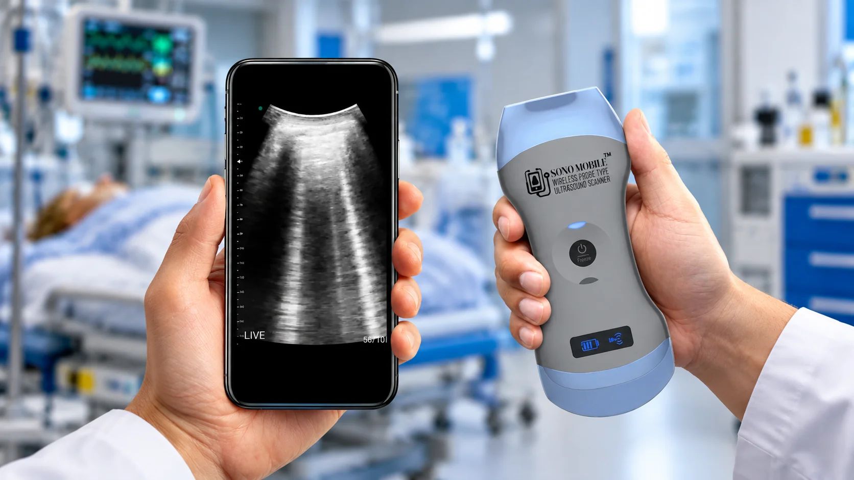
Lung Ultrasound Guide
This practical lung ultrasound guide covers bedside scanning technique, A-lines and B-lines, lung sliding, lung point, normal findings, pneumothorax, pleural effusion, consolidation and common POCUS protocols. It is designed for clinicians and learners who want a structured approach to lung ultrasound POCUS and bedside interpretation.
Why Lung Ultrasound Matters in Point-of-Care Ultrasound
Lung ultrasound is a focused application of Point-of-Care Ultrasound (POCUS) that provides dynamic bedside information without ionizing radiation. Because it can be repeated during reassessment, it can complement the physical examination and other diagnostic testing when clinically appropriate.
In emergency, critical care and pulmonary settings, clinicians may use lung ultrasound to evaluate patients with dyspnea, hypoxemia, chest trauma, suspected pleural fluid, interstitial-alveolar patterns, peripheral consolidation or suspected pneumothorax. For the specialty-focused clinical pathway, see Lung POCUS for Pulmonary Care.

Basic Lung Ultrasound Anatomy and Artifacts
Lung ultrasound differs from ultrasound of solid organs because normally aerated lung contains air. Much of the examination therefore depends on recognizing the pleural interface and characteristic artifacts rather than directly visualizing normal lung parenchyma.
Important structures and signs include the pleural line, rib shadows, lung sliding, horizontal A-lines, vertical artifacts commonly described as B-lines, subpleural consolidation and pleural fluid. These findings should be interpreted as patterns rather than isolated abnormalities.
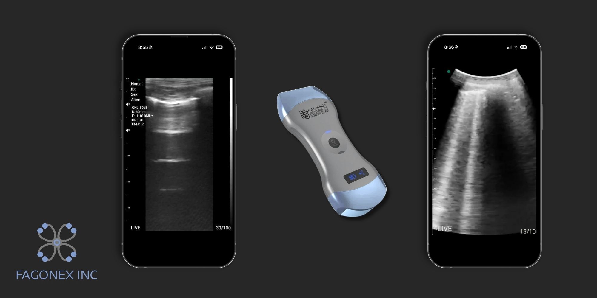
A-Lines vs B-Lines in Lung Ultrasound
A-Lines
A-lines are horizontal reverberation artifacts that repeat beneath the pleural line. In an appropriate scanning window, a predominant A-line pattern is commonly associated with aerated lung.
B-Lines
B-lines are vertical artifacts arising from the pleural line that extend toward the bottom of the image and typically move with respiration. They may partially or completely obscure A-lines.
What Do B-Lines on Lung Ultrasound Mean?
B-lines on lung ultrasound should not be interpreted as a diagnosis by themselves. Their number, morphology and distribution across the chest are important. A small number of isolated B-lines may occur in otherwise aerated lung, particularly in dependent regions.
Multiple, bilateral or diffuse B-lines on lung ultrasound may be seen with increased extravascular lung water and cardiogenic pulmonary edema, while focal B-lines can occur with several localized pulmonary processes. Confluent B-lines describe vertical artifacts that merge as aeration decreases, but their significance still depends on the complete sonographic and clinical pattern.
B-Lines and Pulmonary Edema
In the appropriate clinical context, a diffuse bilateral B-line pattern can support the assessment of pulmonary congestion. However, B-lines are not disease-specific. Integrating lung findings with history, examination and, when appropriate, Cardiac POCUS provides a more complete cardiopulmonary assessment.
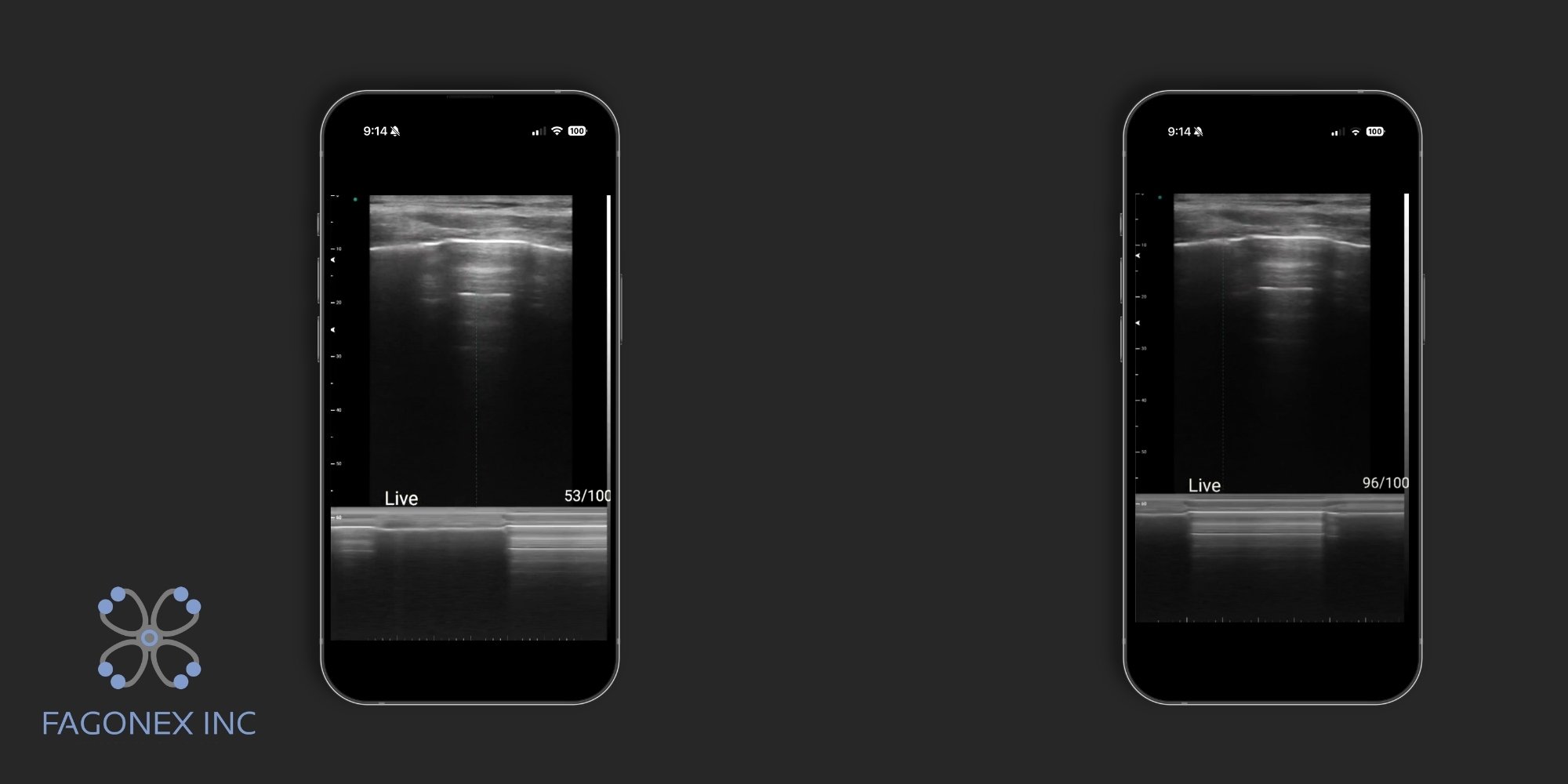
Lung Sliding, Lung Pulse and Lung Point
Lung Sliding
Lung sliding is the shimmering movement of the pleural interface during respiration. When clearly present at the site being examined, it strongly argues against pneumothorax at that location.
On M-mode, normal pleural movement may produce the classic seashore appearance. In contrast, absent lung sliding can produce a barcode or stratosphere pattern. Importantly, absent lung sliding is not specific for pneumothorax and should be interpreted together with other sonographic signs and the clinical situation.
This distinction is particularly important in emergency medicine ultrasound, where rapid bedside assessment still requires systematic interpretation rather than reliance on a single sign.
Lung Pulse
Lung pulse is subtle pleural movement synchronized with cardiac activity rather than respiration. Its presence supports apposition of the pleural layers at the examined site and can be useful when respiratory movement is absent or markedly reduced.
Lung Point POCUS
The lung point describes a transition between an area without normal lung sliding and an area where sliding intermittently reappears with respiration. In the appropriate clinical context, identification of a lung point is a highly specific sonographic sign of pneumothorax.
The sign may not be visualized in every pneumothorax, and its location can vary with pneumothorax size and patient position. Lung point assessment should therefore remain part of a complete focused examination rather than an isolated test.

Lung Ultrasound Probe Selection and Probe Position
Probe choice depends on the clinical question, patient habitus and required depth. Common options include:
- Linear probe: high-resolution visualization of the pleural line and superficial structures.
- Curvilinear probe: broader field and greater depth for pleural fluid, diaphragm visualization and peripheral consolidation.
- Phased-array probe: useful when lung ultrasound is integrated with focused cardiac assessment.
Lung Ultrasound Probe Position
For a basic longitudinal view, place the probe between two ribs with the orientation marker directed according to the convention used by your institution. The ribs and their acoustic shadows frame the pleural line beneath them.
The probe can then be moved systematically through anterior, lateral and posterior lung regions according to the examination protocol and patient position.
A multi-probe workflow is particularly useful when respiratory symptoms require combined pulmonary and cardiac assessment. See our Cardiac POCUS resource for the cardiac component of this clinical cluster.

Step-by-Step Lung Ultrasound Scanning Technique
- Position the patient according to the clinical situation and areas that need examination.
- Select the appropriate probe, depth and lung preset.
- Identify the ribs and pleural line before interpreting artifacts.
- Scan anterior, lateral and posterior regions as clinically appropriate.
- Compare corresponding regions of the right and left lungs.
- Assess lung sliding, A-lines, B-lines, pleural fluid and peripheral consolidation.
- Save representative still images or cine loops and document the examined regions.
Different protocols use different numbers of lung zones. A consistent, systematic approach is more important than speed during early training. Clinicians learning bedside imaging may also benefit from structured handheld ultrasound education and training.

Normal Lung Ultrasound Findings
A typical normal pattern may include a regular pleural interface, visible lung sliding, predominant A-lines and no more than occasional isolated B-lines. Normal findings at the scanned locations do not exclude every pulmonary condition, so the examination should always remain tied to the clinical question.
Pneumothorax on Lung Ultrasound
Pneumothorax lung ultrasound assessment relies on a combination of findings rather than one isolated sign. Clinicians may evaluate lung sliding, B-lines, lung pulse, M-mode patterns and the presence of a lung point.
Absent lung sliding alone is insufficient to establish pneumothorax. When the examination is limited or equivocal, or when comprehensive imaging is clinically required, additional diagnostic imaging should be obtained according to local protocols.

Pleural Effusion Ultrasound
Pleural fluid generally appears as an anechoic or hypoechoic collection above the diaphragm. Depending on the volume and underlying physiology, adjacent atelectatic lung may be visible moving within the fluid. Ultrasound can help identify and localize pleural fluid at the bedside.
Pulmonary Edema and Bilateral B-Lines
Cardiogenic pulmonary edema may produce multiple diffuse, bilateral B-lines. The distribution is often more informative than a single artifact. Because similar vertical artifacts can occur in other conditions, lung findings should be integrated with the overall clinical assessment and, where appropriate, focused cardiac ultrasound.
Lung Consolidation and Pneumonia Patterns
Peripheral consolidation may appear tissue-like or “hepatized” on lung ultrasound. Static or dynamic air bronchograms, pleural irregularity and associated pleural fluid may also be seen.
Lung ultrasound is best able to identify pulmonary abnormalities that reach the pleural surface. Deeper lesions surrounded by normally aerated lung may not be visible, which is an important limitation when interpreting a negative examination.

Lung Ultrasound Protocols: BLUE, eFAST and Bedside Assessment
BLUE Protocol POCUS
The BLUE protocol is a structured lung ultrasound approach developed for patients with acute respiratory failure. It combines predefined lung profiles and clinical pathways to help clinicians differentiate common causes of acute dyspnea. It should be applied by appropriately trained clinicians within its intended clinical context.
eFAST
The extended Focused Assessment with Sonography in Trauma (eFAST) adds thoracic views to the traditional FAST examination. Lung views can contribute to the focused assessment for pneumothorax and pleural fluid in trauma patients.
Bedside and Serial Lung Assessment
Zone-based lung scanning can also support focused reassessment in emergency and critical care settings. Repeat examinations may help clinicians evaluate changes over time when ultrasound findings are integrated with the broader clinical picture.
Common Lung Ultrasound Mistakes and Interpretation Pitfalls
- Using absent lung sliding alone to diagnose pneumothorax.
- Treating B-lines as a disease-specific finding.
- Scanning too few lung regions or failing to compare both sides.
- Confusing other vertical artifacts or subcutaneous findings with pulmonary pathology.
- Using inappropriate depth, gain or probe selection.
- Interpreting ultrasound findings without considering the clinical context.

Documentation, Image Storage and Follow-Up
A focused lung ultrasound examination should document the regions examined and relevant positive or negative findings. Representative still images or cine loops can support comparison, quality review and integration into the clinical record according to institutional policies.
Handheld Ultrasound for Lung POCUS
Handheld ultrasound can bring focused lung imaging to emergency departments, critical care units, clinics and other point-of-care environments. Portability can facilitate bedside imaging and serial reassessment when a conventional cart-based system is not immediately available.
For clinicians who need lung and cardiac imaging in a single portable workflow, Sono Mobile® CL64 combines linear, convex and phased-array scanning capabilities in a wireless handheld system.
Explore Lung POCUS and Handheld Ultrasound
Continue to the pulmonary specialty page for clinical lung POCUS applications, or explore Sono Mobile® CL64 for a multi-organ point-of-care ultrasound workflow.
Conclusion
Lung ultrasound is a practical component of modern point-of-care ultrasound. A systematic examination can help clinicians recognize pleural and lung artifacts, assess common bedside findings and integrate pulmonary information with other focused ultrasound examinations.
This lung ultrasound guide provides an educational framework for technique and interpretation. Continue with Pulmonary Lung POCUS for specialty applications, Cardiac POCUS for integrated cardiopulmonary assessment, and Emergency Medicine Ultrasound for acute-care workflows.