
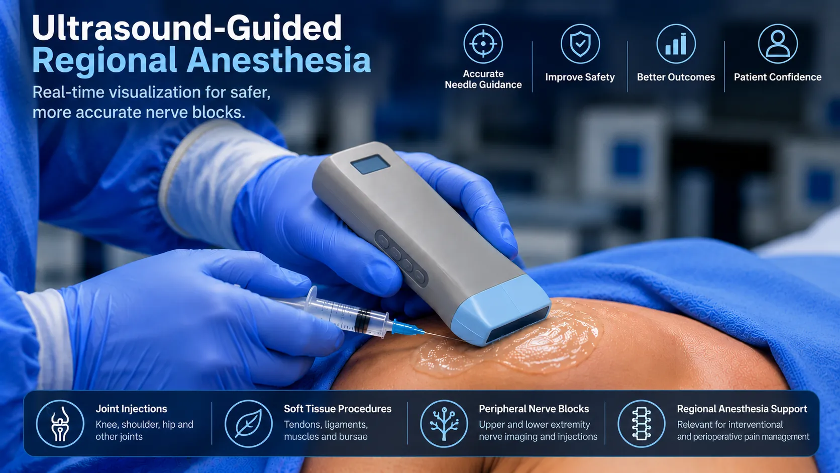
Ultrasound guided regional anesthesia has become an important component of modern perioperative care. By providing real-time visualization of peripheral nerves, surrounding anatomy, and relevant vascular structures, ultrasound can support more informed planning and performance of selected regional anesthesia procedures.
For anesthesiologists and other appropriately trained clinicians, ultrasound guided regional anesthesia offers a way to integrate anatomical imaging into peripheral nerve block practice. However, successful outcomes depend on much more than visualization alone. Clinical assessment, patient selection, anatomical knowledge, aseptic technique, monitoring, and professional training remain essential.
This clinical overview examines the principles, common applications, potential benefits, limitations, and safety considerations of ultrasound guided regional anesthesia. For the broader role of bedside imaging, visit the FAGONEX point-of-care ultrasound (POCUS) guide .
Clinical focus: This article concentrates on regional anesthesia and peripheral nerve blocks. For a broader discussion of joint, tendon, bursal, and other image-guided interventions, read our separate article on ultrasound guided injections .
What Is Ultrasound Guided Regional Anesthesia?
Ultrasound guided regional anesthesia (UGRA) refers to the use of ultrasound imaging to assist in locating relevant anatomical structures during selected regional anesthetic procedures. In peripheral nerve blocks, ultrasound can help clinicians identify nerves or fascial compartments, assess nearby tissues, and observe aspects of needle positioning and local anesthetic distribution in real time.
Regional anesthesia aims to provide anesthesia or analgesia to a defined anatomical region. Depending on the procedure, clinical indication, and patient-specific circumstances, it may be used as part of surgical anesthesia, postoperative pain management, or selected acute pain strategies.
Ultrasound does not replace clinical examination, anatomical knowledge, or established monitoring practices. It is an imaging tool integrated into a comprehensive clinical workflow. Our anesthesia ultrasound specialty page explores additional applications, including vascular access and focused perioperative ultrasound assessment.
How Ultrasound Guidance Supports Peripheral Nerve Blocks
An ultrasound guided nerve block involves using sonographic information to support the identification of relevant neural anatomy and surrounding structures. Depending on the nerve, depth, patient anatomy, and imaging conditions, ultrasound may provide direct or indirect visualization of the intended target.
Peripheral nerves may demonstrate different sonographic appearances depending on their anatomical location and imaging plane. Some exhibit a fascicular pattern, while others may be more difficult to distinguish from nearby tissues. Interpretation therefore requires knowledge of normal anatomy, anatomical variation, and common imaging artifacts.
Anatomical Identification
Ultrasound allows clinicians to assess the relationship between nerves, muscles, fascia, blood vessels, and other surrounding structures. This information may be particularly useful when surface landmarks are difficult to interpret or when individual anatomy differs from expected patterns.
Real-Time Imaging
Dynamic imaging can provide additional information during procedural assessment. The ability to observe anatomy in real time is a distinguishing feature of ultrasound compared with techniques based solely on surface landmarks.
Doppler Assessment
Color or power Doppler imaging may help identify blood flow in nearby vascular structures when clinically appropriate. Doppler information should be interpreted alongside grayscale imaging and clinical anatomy, since technical settings and vessel characteristics can influence flow detection.
Common Applications of Ultrasound Guided Regional Anesthesia
Ultrasound guided peripheral nerve block techniques are used across a range of anatomical regions. The selection of a block depends on the surgical or clinical indication, expected sensory distribution, patient factors, contraindications, and local expertise.
Upper-Extremity Nerve Blocks
Upper-extremity regional anesthesia may involve selected approaches to the brachial plexus or individual peripheral nerves. Ultrasound can assist in assessing neural anatomy and adjacent vascular structures relevant to the planned procedure.
Femoral and Lower-Extremity Blocks
An ultrasound guided femoral nerve block is one example of lower-extremity regional anesthesia. Other approaches may target selected nerves or fascial compartments according to the clinical indication and anticipated functional effects.
Fascial Plane Blocks
Fascial plane blocks involve the administration of local anesthetic within selected tissue planes. Ultrasound can help identify relevant muscular and fascial anatomy, although effectiveness and evidence vary between techniques and indications.
Selected Acute Pain Applications
Regional techniques may contribute to multimodal analgesia in selected acute pain settings. Their use requires consideration of the clinical environment, available monitoring, practitioner competence, and follow-up arrangements.
Regional techniques also intersect with broader clinical areas such as ultrasound in pain management and musculoskeletal ultrasound . These related disciplines share anatomical imaging principles but differ in clinical objectives, procedural indications, and patient-management requirements.
Benefits of Ultrasound Guided Regional Anesthesia
Published evidence suggests that ultrasound guidance can improve selected characteristics of regional anesthetic blocks. The magnitude and clinical relevance of these benefits vary by block type, comparator technique, operator experience, and patient population.
- Improved anatomical visualization: Ultrasound can help identify relevant nerves, tissue planes, and nearby anatomical structures.
- Support for procedural planning: Imaging provides information that may influence the selection and assessment of an appropriate regional technique.
- Selected improvements in block characteristics: Evidence supports improvements in certain performance and success measures, although effects vary among different blocks.
- Potential reduction of selected complications: Evidence indicates benefits for certain adverse outcomes, including local anesthetic systemic toxicity, but ultrasound does not eliminate procedural risks.
- Integration with multimodal care: Appropriate regional techniques may support perioperative analgesia plans when used within established clinical protocols.
An evidence-based assessment published in Regional Anesthesia & Pain Medicine found that ultrasound guidance offers advantages for selected blocks, while emphasizing that improvements are not uniform across all outcomes. See the ASRA evidence-based assessment for further discussion.
Limitations and Clinical Challenges
Despite its advantages, ultrasound guidance has important limitations. Image quality can be affected by tissue depth, body habitus, acoustic windows, equipment characteristics, and operator technique. Some neural structures may be difficult to visualize consistently.
Ultrasound images also require accurate interpretation. Artifacts, anisotropy, and similarities between neural and non-neural tissues can complicate anatomical identification. Furthermore, visualization of a needle or surrounding anatomy does not guarantee correct placement or prevent injury.
Importantly, available evidence has not established that ultrasound guidance eliminates postoperative neurological complications. Regional anesthesia remains an invasive clinical intervention requiring appropriate training, monitoring, and emergency preparedness.
Patient Safety in Ultrasound Guided Regional Anesthesia
Patient safety is central to the appropriate use of ultrasound guided regional anesthesia. Imaging should be considered one component of a broader clinical safety system, rather than a substitute for established procedural safeguards.
Patient Selection and Clinical Assessment
Before a regional technique is considered, clinicians must assess the indication, relevant medical history, neurological status, potential contraindications, patient preferences, and the expected benefits and risks. Anticoagulant therapy, infection risk, and other patient-specific considerations may affect procedural planning.
Aseptic Technique and Infection Prevention
Infection prevention includes appropriate hand hygiene, skin preparation, ultrasound equipment cleaning, and the use of suitable sterile supplies according to the procedure and current guidance.
ASRA Pain Medicine's infection control guidance addresses probe covers, sterile gel, disinfection, and infection prevention during regional anesthesia and pain interventions. See the ASRA infection control guidelines .
Monitoring and Complication Readiness
Appropriate patient monitoring, communication, documentation, and access to emergency management resources are essential. Complications may include local anesthetic systemic toxicity, nerve injury, bleeding, infection, or other block-specific adverse events. Ultrasound guidance does not remove the need to recognize and manage these risks.
Safety reminder: Ultrasound guided regional anesthesia should be performed only by appropriately qualified and trained healthcare professionals, within their scope of practice and institutional protocols. This article is educational and does not provide a procedural protocol, drug dosing instructions, or patient-specific medical advice.
Choosing an Ultrasound System for Regional Anesthesia
Ultrasound equipment selection depends on the intended anatomical targets, required imaging depth, probe configuration, image quality, display characteristics, and clinical workflow. There is no single probe configuration that is optimal for every regional anesthesia application.
| Consideration | Clinical Relevance |
|---|---|
| Linear Transducer | Commonly used for many relatively superficial peripheral nerve and vascular applications. |
| Curved Transducer | May be useful when deeper anatomical structures require a wider field of view. |
| Image Optimization | Depth, gain, focus, and suitable presets influence anatomical interpretation. |
| Doppler Imaging | Can support assessment of relevant vascular structures when appropriately applied. |
| Portability | May support bedside, operating-room, educational, and mobile workflows. |
| Cleaning and Infection Control | Equipment must be compatible with appropriate disinfection and sterile-cover protocols. |
Handheld Ultrasound for Regional Anesthesia Workflows
Portable and handheld ultrasound systems can make anatomical imaging available in settings where a larger cart-based system may be less convenient. Wireless configurations may support flexible positioning and educational demonstrations, provided the device meets the required clinical and infection-control standards.
The Sono Mobile® CL64 handheld ultrasound combines linear and convex transducer configurations with a phased-array preset in a wireless dual-head system. It provides B-mode, M-mode, Color Doppler, Power Doppler, and Pulsed-Wave Doppler imaging capabilities.
For appropriately trained clinicians, the CL64 may support selected regional anesthesia imaging tasks alongside other POCUS applications. Suitability for a specific procedure should be assessed according to imaging requirements, operator competence, local regulations, and clinical protocols.
Explore the broader range of Sono Mobile® handheld ultrasound systems to compare probe configurations and available features.
Training and Competency in Ultrasound Guided Regional Anesthesia
Effective ultrasound guided regional anesthesia requires more than familiarity with ultrasound equipment. Training includes understanding sonoanatomy, recognizing anatomical variation, interpreting imaging artifacts, maintaining procedural safety, and applying appropriate clinical decision-making.
Structured education, supervised practice, competency assessment, and ongoing quality improvement are important components of a responsible clinical program. Educational requirements vary according to professional role, jurisdiction, and institutional policy.
Broader foundational concepts are discussed in our POCUS educational guide , while the medical ultrasound specialties hub connects readers to additional clinical applications.
Frequently Asked Questions
What is ultrasound guided regional anesthesia?
Ultrasound guided regional anesthesia uses ultrasound imaging to assist clinicians in identifying relevant anatomy during selected regional anesthetic procedures, including peripheral nerve blocks.
What are the benefits of an ultrasound guided nerve block?
Potential benefits include improved anatomical visualization and selected improvements in block performance characteristics. Benefits vary by technique, clinical indication, and operator experience.
Can ultrasound eliminate nerve block complications?
No. Ultrasound may reduce certain risks, but it cannot eliminate complications such as neurological injury, bleeding, infection, or local anesthetic systemic toxicity.
Which ultrasound probe is used for peripheral nerve blocks?
Linear transducers are commonly used for many superficial applications, while curved transducers may be considered for deeper targets. Selection depends on the anatomy, required imaging depth, and clinical context.
Can handheld ultrasound be used for regional anesthesia?
Selected handheld systems may support regional anesthesia imaging when their capabilities are appropriate for the intended procedure and they are used by trained clinicians following established safety protocols.
Conclusion
Ultrasound guided regional anesthesia integrates real-time anatomical imaging into selected peripheral nerve block and regional anesthetic workflows. Its value lies in supporting anatomical assessment and informed clinical practice, while recognizing that imaging alone cannot guarantee procedural success or patient safety.
As ultrasound becomes increasingly integrated into perioperative care, appropriate equipment, professional training, evidence-based protocols, and ongoing competency assessment remain essential. Explore our anesthesia POCUS resources for related clinical applications.
Explore Sono Mobile® for Anesthesia POCUS
Discover how portable ultrasound imaging may support selected anesthesia and perioperative workflows. Review the Sono Mobile® CL64 specifications or contact FAGONEX to discuss device features, compatibility, and demonstration options.
Scientific References
- Neal JM, et al. The Second American Society of Regional Anesthesia and Pain Medicine Evidence-Based Medicine Assessment of Ultrasound Guided Regional Anesthesia: Executive Summary. Regional Anesthesia & Pain Medicine. PubMed .
- ASRA Pain Medicine. Consensus Practice Infection Control Guidelines for Regional Anesthesia and Pain Medicine. Regional Anesthesia & Pain Medicine. Clinical Guidelines .
- ASRA Practice Advisory on Neurologic Complications in Regional Anesthesia and Pain Medicine. Full Text .
- Ultrasound Guided Regional Anesthesia and Patient Safety: Update of an Evidence-Based Analysis. PubMed .
Medical Disclaimer: This material is intended for professional education and general informational purposes. It does not replace clinical training, institutional protocols, professional guidelines, or individualized medical judgment. Clinical procedures should be undertaken only by qualified healthcare professionals within their authorized scope of practice.