
Musculoskeletal Ultrasound Scanning: A Practical Guide to Probe Positioning and Imaging Techniques
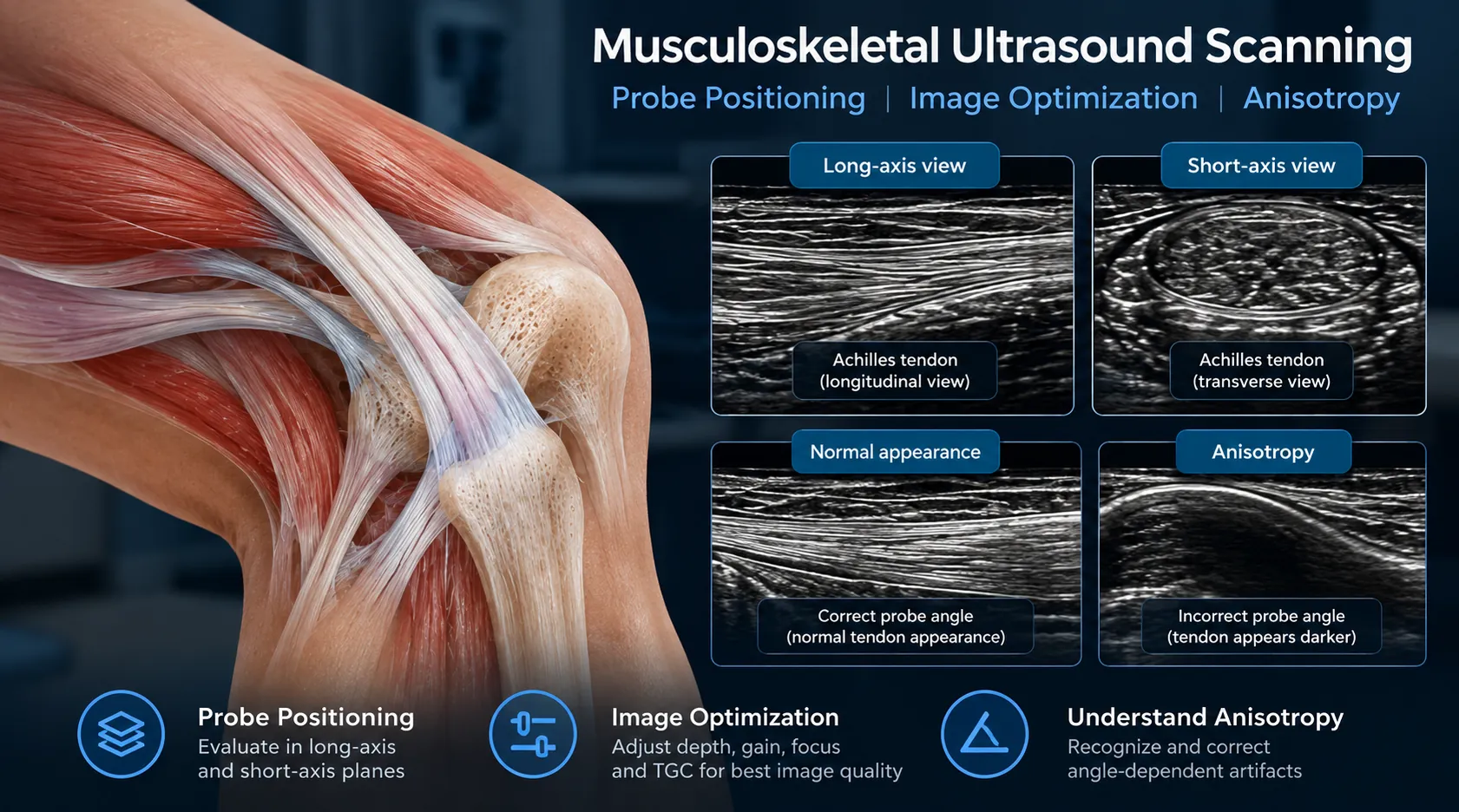
Effective musculoskeletal ultrasound scanning depends on more than image quality. Transducer selection, patient positioning, scanning planes, probe orientation, and artifact recognition all influence how muscles, tendons, ligaments, and superficial joints appear on ultrasound.
Proper ultrasound probe positioning is essential for accurate musculoskeletal scanning. It helps clinicians obtain consistent long-axis and short-axis views while minimizing imaging artifacts such as anisotropy.
This practical guide introduces essential MSK scanning techniques for healthcare professionals. For an overview of clinical applications, visit the MSK ultrasound specialty guide.
Effective ultrasound probe positioning is essential for obtaining consistent long-axis and short-axis views during musculoskeletal examinations.
1. Choosing the Right Ultrasound Transducer
High-frequency linear transducers are commonly used for superficial musculoskeletal structures because they provide detailed visualization of tendons, muscles, ligaments, peripheral nerves, and superficial soft tissues.
Lower-frequency settings or alternative transducers may be required for deeper structures. Selection should reflect the clinical question, patient anatomy, imaging depth, and available equipment.
| Consideration | Practical approach |
|---|---|
| Superficial tendons | Start with an appropriate high-frequency linear probe. |
| Deeper muscles | Adjust frequency and penetration to the required depth. |
| Small structures | Optimize depth, focal zone, and probe contact. |
| Dynamic assessment | Maintain a stable image while performing controlled movement. |
The AIUM musculoskeletal ultrasound practice parameter provides professional guidance on examination technique, documentation, and clinical considerations.
2. Patient Positioning and Examination Preparation
Patient positioning should provide comfortable access to the anatomical region while minimizing unnecessary muscle tension. A supported and reproducible position helps clinicians obtain consistent longitudinal and transverse views.
- Identify the clinical indication and target anatomy.
- Position the patient to expose the examination region.
- Use sufficient coupling gel for continuous acoustic contact.
- Locate reliable anatomical landmarks before detailed scanning.
- Compare with the contralateral side when clinically useful.
- Document orientation, location, and relevant findings.
3. Long-Axis vs. Short-Axis MSK Scanning
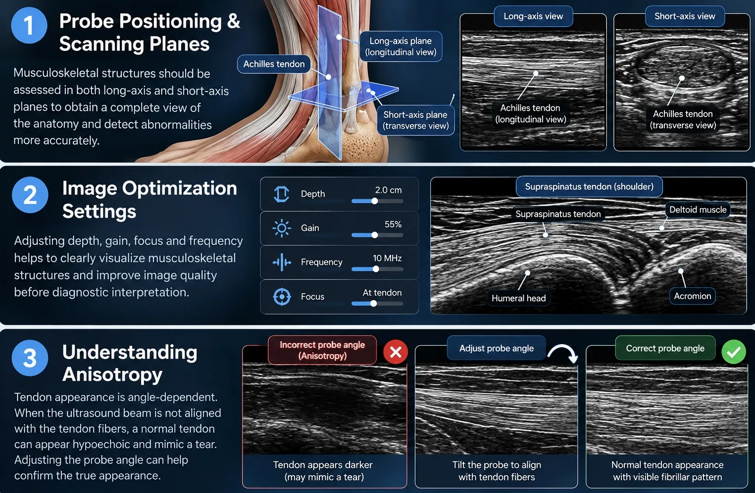
Most elongated musculoskeletal structures should be assessed in at least two complementary planes. Scanning in only one plane can obscure abnormalities or create misleading appearances.
Long-Axis Scanning
Align the imaging plane along the structure's length. This helps demonstrate tendon fibrillar patterns, muscle fiber direction, and longitudinal continuity.
Short-Axis Scanning
Rotate the probe to examine the structure in cross-section. This can help identify boundaries, adjacent anatomy, and the transverse extent of a finding.
4. Ultrasound Probe Positioning and Movements
Proper ultrasound probe positioning involves controlled sliding, tilting, rocking, and rotation to maintain anatomical alignment and improve image interpretation.
Probe manipulation is a fundamental component of musculoskeletal ultrasound scanning. Small adjustments can substantially change the visibility of tendons, muscles, and other superficial structures.
Sliding
Move the transducer across the skin to follow anatomy or identify a nearby structure.
Tilting
Change the beam angle to improve alignment with the target and reduce angle-dependent artifacts.
Rocking
Adjust the probe around its contact area to maintain an appropriate insonation angle along curved anatomy.
Rotation
Turn the transducer to transition between longitudinal and transverse views.
The EULAR standardized scanning framework emphasizes consistent probe placement, anatomical landmarks, and structured examination techniques. Read the EULAR scanning procedures publication .
5. Optimizing MSK Ultrasound Images
Image optimization should occur before diagnostic interpretation. A technically inadequate image can reduce confidence in the appearance of otherwise accessible anatomy.
| Setting | Why it matters |
|---|---|
| Depth | Keep the target and relevant surrounding anatomy visible. |
| Gain | Balance tissue brightness without obscuring detail. |
| Frequency | Balance superficial resolution with required penetration. |
| Focus | Position the focal region appropriately for the target. |
| Probe pressure | Avoid unnecessarily compressing small fluid collections or vessels. |
| Doppler settings | Optimize sensitivity and reduce motion artifacts when vascularity is relevant. |
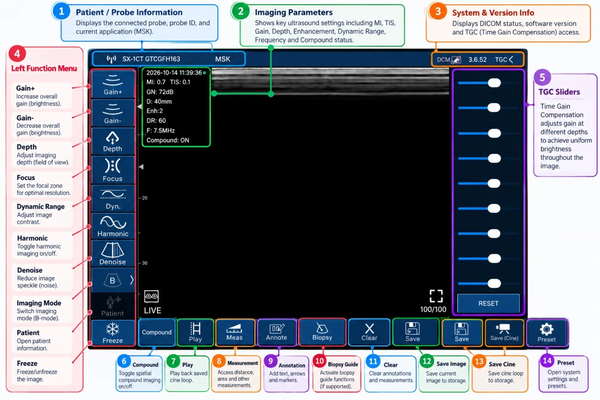
Image quality also depends on stable hand positioning, appropriate acoustic coupling, and recognition of normal anatomical variation.
6. Understanding Anisotropy and Common Artifacts
Anisotropy is especially important in tendon ultrasound. When the sound beam is not appropriately aligned with tendon fibers, a normal tendon may appear artificially hypoechoic and resemble pathology.
To evaluate suspected anisotropy, adjust the transducer angle and reassess the structure in multiple planes. Persistent abnormality after appropriate correction requires clinical interpretation rather than an automatic assumption of injury.
Accurate ultrasound probe positioning helps minimize anisotropy by maintaining an appropriate insonation angle relative to tendon fibers.
Common Scanning Pitfalls
- Interpreting anisotropy as a tendon tear.
- Using excessive imaging depth for a superficial target.
- Applying too much pressure over small fluid collections.
- Failing to assess a structure in two planes.
- Confusing normal anatomical variants with abnormalities.
- Using motion-sensitive Doppler settings without adequate stabilization.
Correct probe positioning and recognition of anisotropy are important when evaluating suspected muscle injuries. For a more detailed discussion of sonographic findings and injury grading, read our muscle tear ultrasound diagnosis and grading guide .
7. Practical MSK Ultrasound Scanning Examples
Shoulder Ultrasound
Shoulder examinations commonly involve the rotator cuff, long head of the biceps tendon, and selected periarticular structures. Consistent positioning and careful probe orientation are particularly important for curved tendons.
Knee Ultrasound
Anterior knee scanning may demonstrate the quadriceps tendon, patellar tendon, suprapatellar recess, and surrounding superficial anatomy. Longitudinal and transverse views provide complementary information.
Muscle Scanning
Muscle ultrasound requires recognition of normal fascicular architecture and comparison across scanning planes. Suspected muscle tears require assessment of fiber continuity, fluid collections, and clinical context.
For detailed injury-related findings, read our Muscle Tear Ultrasound: Imaging Findings, Diagnosis & Grading article.
8. A Practical MSK Scanning Checklist
- Confirm the indication and target anatomy.
- Select an appropriate transducer and preset.
- Position the patient and identify landmarks.
- Adjust depth, gain, frequency, and focus.
- Scan systematically in long and short axes.
- Correct anisotropy before interpreting tendon appearance.
- Use controlled dynamic assessment when appropriate.
- Record representative images and relevant findings.
- Recognize examination limitations and when further imaging is needed.
9. Handheld Ultrasound for MSK Scanning
Wireless handheld ultrasound can support selected musculoskeletal examinations in clinics, educational settings, and point-of-care environments. Device selection should be based on image performance, transducer configuration, workflow, and the intended clinical task.
The Sono Mobile® CL64 combines convex and linear scanning capabilities in a dual-head device. Its linear frequencies of 7.5 and 10 MHz may support selected superficial MSK applications.
The Sono Mobile® L4 provides a dedicated wireless linear transducer with 7.5 and 10 MHz settings for selected superficial imaging applications.
Explore the Sono Mobile® handheld ultrasound range or learn how these devices fit within point-of-care ultrasound workflows .
Explore Wireless Handheld Ultrasound for MSK Imaging
Compare dual-head and dedicated linear ultrasound configurations for your clinical and educational needs. Discuss your scanning workflow with the FAGONEX team.
10. Frequently Asked Questions
Which probe is best for musculoskeletal ultrasound?
A high-frequency linear transducer is commonly used for superficial MSK anatomy. Deeper structures may require different frequency or transducer settings.
Why scan in both long and short axes?
Orthogonal imaging helps evaluate structural continuity, anatomical relationships, and the extent of findings. It also reduces the risk of misinterpretation from a single imaging plane.
What is anisotropy in tendon ultrasound?
Anisotropy is an angle-dependent change in echogenicity. It can make normal tendon tissue appear abnormally dark when the beam is not properly aligned.
Can handheld ultrasound be used for MSK examinations?
Yes, appropriately configured handheld devices can support selected MSK examinations. Clinical suitability depends on anatomy, image quality, operator training, and the examination requirements.
Can ultrasound replace MRI for all MSK injuries?
No. Ultrasound is useful for many accessible superficial structures and dynamic examinations, while MRI may be preferable for deeper or more complex clinical questions.
Scientific References & Further Reading
- American Institute of Ultrasound in Medicine. The AIUM Practice Parameter for the Performance of the Musculoskeletal Ultrasound Examination. Journal of Ultrasound in Medicine. 2023;42:E23–E35. DOI: 10.1002/jum.16228. PubMed →
- AIUM. Musculoskeletal Ultrasound Practice Parameters. Official AIUM resource →
- EULAR. The 2017 EULAR Standardised Procedures for Ultrasound Imaging in Rheumatology. Scientific publication →