

Cancer detection has evolved significantly over the last decades, and medical imaging plays a central role in early diagnosis and clinical decision-making. Among imaging modalities, ultrasound stands out as a safe, radiation-free, and highly accessible technology. But a common question remains: Can ultrasound detect cancer?
The short answer is: Ultrasound can help detect many types of tumors and suspicious masses, but it does not diagnose cancer on its own. It is often used as a first-line imaging tool or as part of a broader diagnostic pathway that may include CT, MRI, mammography, laboratory tests, and biopsy.
At Fagonex; Manufacturer of Sono Mobile™ handheld ultrasound systems, we support clinicians worldwide with advanced portable ultrasound solutions designed for point-of-care diagnostics, rapid assessment, and real-time imaging in hospitals, clinics, and remote settings.
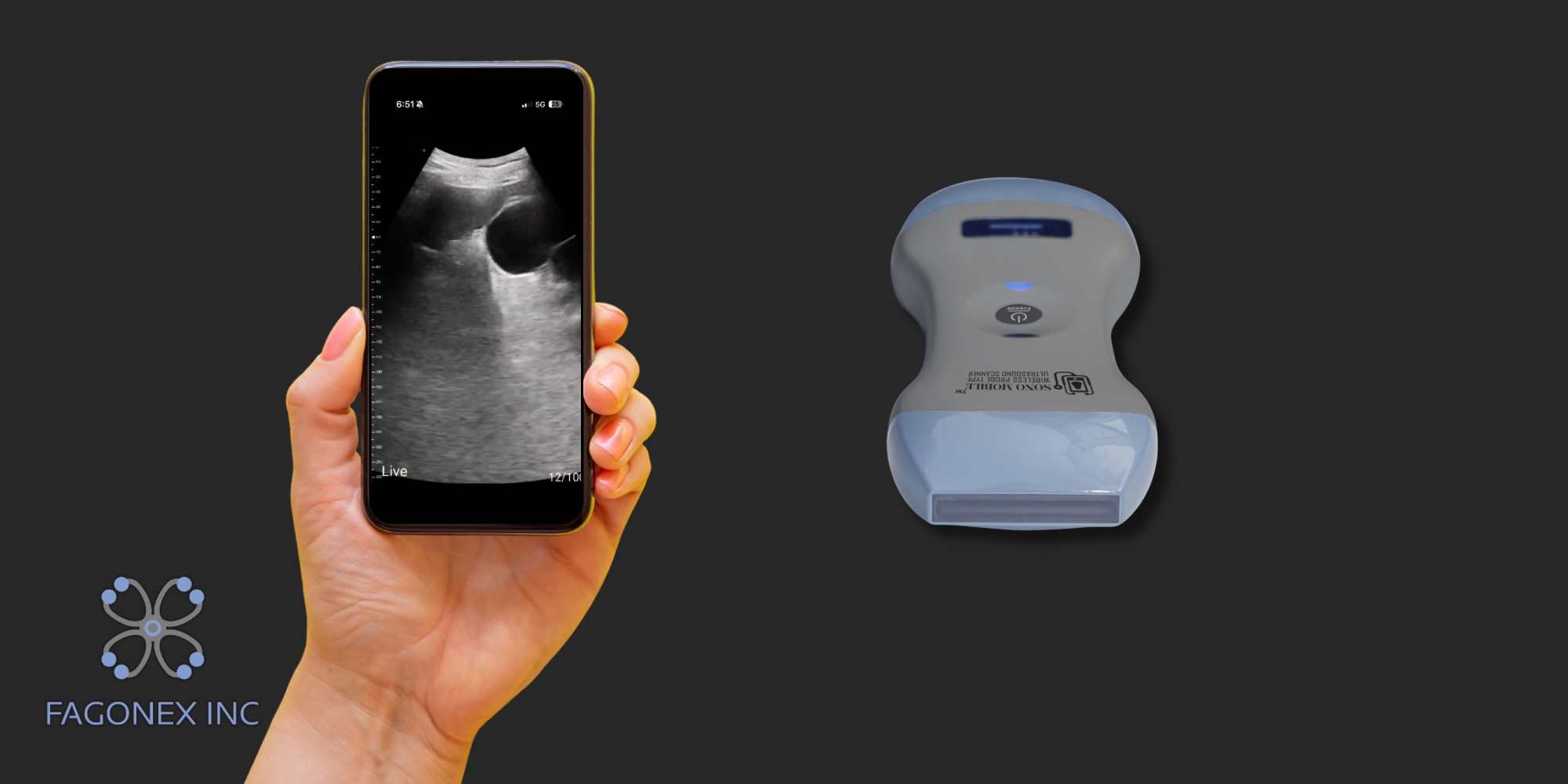
Can Ultrasound Detect Tumors in the Abdomen?
Abdominal ultrasound is one of the most commonly performed diagnostic imaging procedures. It can detect:
- Liver masses
- Pancreatic lesions
- Kidney tumors
- Bladder abnormalities
- Ovarian cysts and masses
- Enlarged lymph nodes
- Fluid collections
Ultrasound identifies abnormal tissue density, irregular borders, solid versus cystic masses, and vascular patterns using Doppler imaging. While it cannot confirm malignancy alone, it plays a crucial role in identifying suspicious findings that require further evaluation.
In many healthcare systems, abdominal ultrasound is the first imaging step when patients present with abdominal pain, unexplained weight loss, jaundice, or abnormal laboratory results.
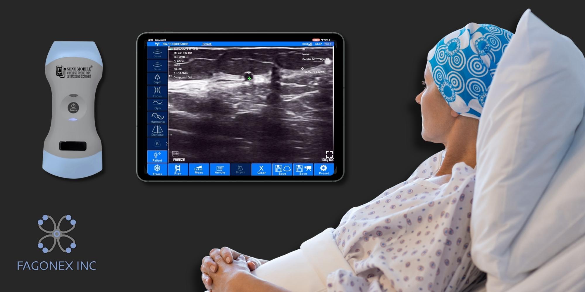
Can Ultrasound Detect Cancer?
Ultrasound can detect masses and structural abnormalities that may represent cancer. However:
- It does not replace biopsy.
- It cannot determine cellular characteristics.
- Some deep or air-filled structures may limit visualization.
Ultrasound is particularly effective in:
- Soft tissue organs
- Fluid-filled structures
- Superficial tumors
- Guiding needle biopsies
Modern portable systems like Sono Mobile™ by Fagonex enhance early detection capabilities by providing real-time, high-resolution imaging at the bedside.

What Cancers Do Not Show Up on Ultrasound?
Ultrasound has limitations. Certain cancers are difficult to visualize, including:
- Early-stage colon cancer
- Lung cancer (air interferes with sound waves)
- Bone cancers
- Very small pancreatic tumors
- Deep brain tumors
This is because ultrasound waves do not travel well through air or bone. In such cases, physicians rely on CT, MRI, PET scans, or endoscopy.
Understanding these limitations is critical for clinicians when choosing the appropriate diagnostic pathway.

How Does Ultrasound Work for Cancer?
Ultrasound uses high-frequency sound waves transmitted through a transducer. These waves reflect differently depending on tissue density and composition.
Key principles in oncology imaging include:
- Hypoechoic vs. hyperechoic patterns
- Irregular margins
- Increased vascularity (via Doppler)
- Tissue stiffness (with elastography)
In cancer evaluation, ultrasound is frequently used to:
Detect Suspicious Masses
Ultrasound plays a critical role in detecting suspicious masses in soft tissues and internal organs. Using high-frequency sound waves, clinicians can identify abnormal growths in the liver, breast, thyroid, ovaries, bladder, and other structures. Suspicious features such as irregular borders, heterogeneous echotexture, hypoechoic patterns, and increased vascularity on Doppler imaging may raise concern for malignancy. As a radiation-free and non-invasive diagnostic imaging modality, ultrasound is often the first-line screening tool in oncology. Early detection of abnormal masses improves clinical outcomes by enabling faster referral for advanced imaging, biopsy, and specialized cancer management pathways.
Monitor Tumor Size
Ultrasound is widely used to monitor tumor size over time, especially in patients undergoing cancer treatment or surveillance programs. By providing real-time imaging, clinicians can measure lesion dimensions accurately and compare them with previous scans. Tracking tumor growth or shrinkage is essential for evaluating disease progression, recurrence, or stability. Portable and point-of-care ultrasound devices enhance follow-up efficiency by allowing bedside reassessment without radiation exposure. In oncology care, consistent tumor measurement supports evidence-based treatment planning, helps adjust therapeutic strategies, and contributes to personalized cancer management protocols focused on improving patient outcomes.
Guide Biopsy Needles
One of the most important applications of ultrasound in oncology is guiding biopsy needles with precision and safety. Ultrasound-guided biopsy allows clinicians to visualize the needle in real time as it enters a suspicious lesion, ensuring accurate tissue sampling. This technique reduces complications, improves diagnostic accuracy, and minimizes damage to surrounding structures. It is commonly used for breast, liver, thyroid, lymph node, and soft tissue biopsies. Compared to blind procedures, ultrasound guidance increases confidence in obtaining representative samples for histopathological analysis, which remains the gold standard for confirming cancer diagnosis and determining appropriate treatment options.
Assess Treatment Response
Ultrasound is an effective imaging tool for assessing treatment response in cancer patients undergoing chemotherapy, radiotherapy, immunotherapy, or targeted therapy. By measuring changes in tumor size, vascularity, and tissue characteristics, clinicians can evaluate whether a treatment plan is working. Doppler ultrasound can reveal reduced blood flow within tumors, while elastography may demonstrate changes in tissue stiffness. Early identification of treatment response or resistance allows oncologists to modify therapeutic strategies promptly. As a safe, repeatable, and cost-effective imaging modality, ultrasound supports continuous monitoring and plays a valuable role in modern, patient-centered oncology care.
Portable ultrasound devices have significantly improved access to cancer screening, particularly in remote and resource-limited environments.

Detecting Bladder Cancer with Ultrasound
Ultrasound can detect:
- Bladder wall thickening
- Intraluminal masses
- Obstruction
- Residual urine volume
While cystoscopy remains the gold standard, ultrasound is a non-invasive initial screening tool often used in urology clinics.
It is especially useful when patients present with:
- Hematuria (blood in urine)
- Urinary retention
- Recurrent infections

Ultrasound in Skin Cancer
High-frequency ultrasound (HFUS) can evaluate:
- Tumor depth
- Margins
- Subcutaneous invasion
- Lymph node involvement
In dermatology, ultrasound assists in staging conditions such as melanoma and non-melanoma skin cancers. It helps guide surgical planning and monitor recurrence.

Ultrasound in Breast Cancer
Evaluate Palpable Lumps
Breast ultrasound is a highly effective imaging modality for evaluating palpable breast lumps detected during physical examination. When a patient or clinician feels a mass, ultrasound provides real-time visualization to determine its size, shape, margins, and internal characteristics. This non-invasive, radiation-free diagnostic tool is especially valuable in younger women and pregnant patients. Ultrasound helps identify suspicious features such as irregular borders, heterogeneous echotexture, and increased vascularity, which may require further investigation. Early assessment of palpable breast lumps improves diagnostic accuracy, supports timely referral for biopsy if needed, and enhances overall breast cancer detection strategies.
Differentiate Cysts from Solid Masses
One of the primary advantages of breast ultrasound is its ability to differentiate cysts from solid masses. Simple cysts appear as well-defined, anechoic (fluid-filled) structures with posterior acoustic enhancement, while solid tumors display internal echoes and variable vascular patterns. This distinction is crucial in breast imaging because many cysts are benign and require only monitoring, whereas solid lesions may need biopsy. By accurately characterizing breast abnormalities, ultrasound reduces unnecessary invasive procedures and improves clinical decision-making. In modern breast cancer screening protocols, ultrasound plays a complementary role alongside mammography to increase diagnostic confidence and patient reassurance.
Assess Dense Breast Tissue
Dense breast tissue can reduce the sensitivity of mammography, making it more challenging to detect early-stage breast cancer. Breast ultrasound is particularly beneficial in women with dense breasts because it provides better visualization of soft tissue structures without relying on X-ray contrast. Supplemental ultrasound screening can detect small, mammographically occult tumors that may otherwise go unnoticed. As awareness about dense breast tissue increases, integrating ultrasound into breast cancer screening programs enhances early detection rates. Portable and high-resolution ultrasound systems further improve accessibility, allowing clinicians to perform comprehensive breast imaging in outpatient and point-of-care settings.
Guide Biopsy
Ultrasound-guided breast biopsy is a precise and minimally invasive procedure used to obtain tissue samples from suspicious lesions. Real-time imaging allows clinicians to accurately position the biopsy needle within the targeted mass, reducing complications and improving diagnostic yield. Compared to blind biopsy techniques, ultrasound guidance enhances safety, shortens procedure time, and increases patient comfort. It is commonly used for sampling solid masses, suspicious lymph nodes, and indeterminate findings identified on mammography or MRI. Accurate tissue diagnosis remains the gold standard for confirming breast cancer, and ultrasound guidance plays a critical role in modern, evidence-based breast care management.
It is particularly valuable in younger women with dense breasts, where mammography sensitivity may be reduced.

When Would You Need a Mammogram and a Breast Ultrasound?
A mammogram is typically recommended:
- For routine screening (age-based guidelines)
- For detecting microcalcifications
- For early detection in asymptomatic women
Breast ultrasound is often used:
- As a complementary test after mammogram
- For evaluating specific areas of concern
- For dense breast tissue
- During pregnancy (radiation-free option)
The combination improves diagnostic accuracy.
For more information on this, be sure to read this article: Breast Ultrasound vs Mammogram
Would You Ever Need a Breast Ultrasound Instead of a Mammogram?
Yes, in certain situations:
- Young patients under screening age
- Pregnant or breastfeeding women
- When evaluating a specific lump
- In resource-limited settings
Portable ultrasound devices enable rapid bedside evaluation and immediate clinical decision-making.
Frequently Asked Question
Can Abdominal Ultrasound Detect Bowel Problems?
Ultrasound can detect:
- Bowel wall thickening
- Obstruction
- Inflammatory changes
- Masses in advanced cases
However, colonoscopy and CT are more sensitive for early colon cancer detection.
Can Ultrasound Detect Cancer in Uterus?
Yes, transvaginal ultrasound can detect:
- Endometrial thickening
- Uterine masses
- Fibroids
- Suspicious lesions
It is commonly used in gynecology as a first-line evaluation.
Can Ultrasound Detect Cancer in Pancreas?
Ultrasound may detect larger pancreatic tumors, but:
- Small lesions are harder to visualize
- Gas in intestines may limit clarity
CT and MRI are often required for confirmation.
Can Ultrasound Detect Cancer in Ovaries?
Yes. Ultrasound is the primary imaging tool for evaluating ovarian masses. It can assess:
- Cystic vs. solid structure
- Septations
- Papillary projections
- Blood flow patterns
Early detection significantly improves outcomes.
Can Ultrasound Detect Cancer in Colon?
Ultrasound has limited sensitivity for early colon cancer. It may detect:
- Large masses
- Obstruction
- Advanced disease
Colonoscopy remains the gold standard.
Can Ultrasound Detect Cancer in Throat?
Ultrasound can assess:
- Thyroid nodules
- Lymph nodes
- Soft tissue masses
For deeper throat cancers, CT, MRI, or endoscopy is preferred.
Can Ultrasound Detect Cancer in Cervix?
Transvaginal ultrasound may identify cervical masses, but Pap smear and HPV testing remain primary screening tools.
Can Ultrasound Detect Cancer in Liver?
Ultrasound is highly effective for liver imaging. It can detect:
- Solid masses
- Cysts
- Cirrhosis-related nodules
- Metastases
It is commonly used for liver cancer screening in high-risk patients.
The Role of Portable Ultrasound in Early Cancer Detection
Modern healthcare increasingly relies on Point-of-Care Ultrasound (POCUS). Portable systems allow:
- Immediate bedside evaluation
- Faster referral decisions
- Reduced waiting times
- Cost-effective screening
- Improved access in rural or developing regions
Sono Mobile™ by Fagonex represents the next generation of handheld ultrasound technology, combining:
- High-resolution imaging
- Doppler capability
- Lightweight design (~250gr)
- Wireless connectivity
- Dual head (convex + linear + cardiac preset)
- Easy sterilization
- Subscription free
For oncologic screening and soft-tissue evaluation, mobility and accessibility can make a meaningful difference in patient pathways.
Conclusion
So, can ultrasound detect cancer?
Ultrasound can detect many tumors and suspicious masses, especially in soft tissues like the liver, breast, ovaries, bladder, and thyroid. While it does not replace biopsy or advanced imaging in all cases, it remains one of the most important and accessible diagnostic tools in modern medicine.
At Fagonex, we are committed to advancing diagnostic imaging through innovative portable ultrasound systems that empower clinicians to make faster, informed decisions; anytime, anywhere.