
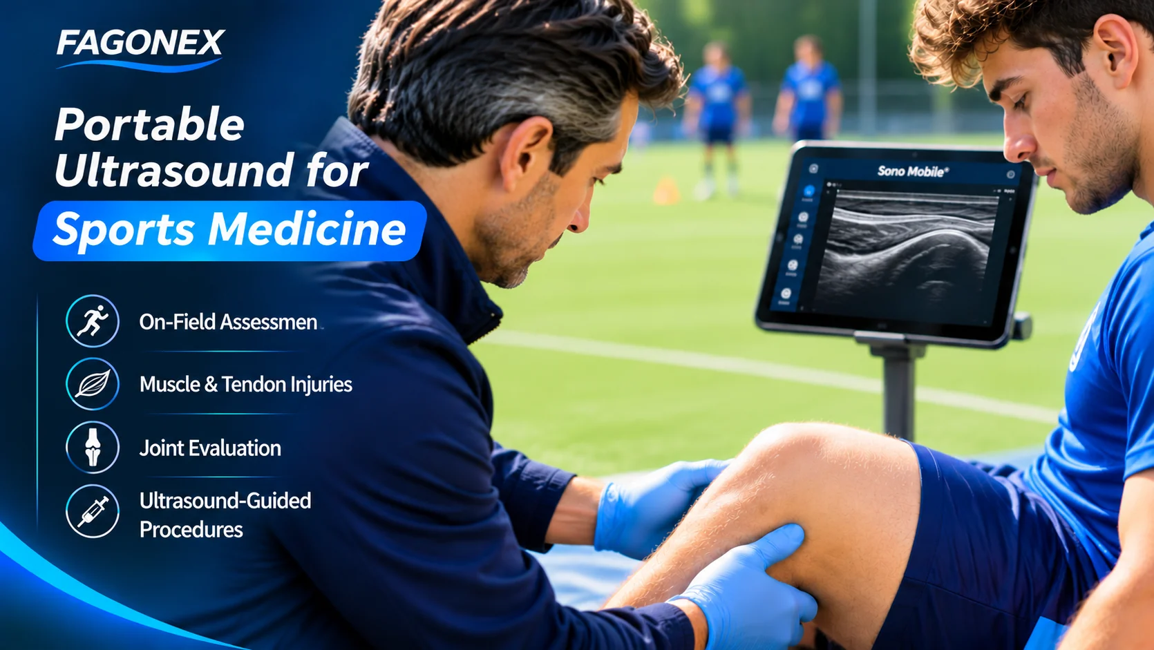
Portable ultrasound for sports medicine brings real-time musculoskeletal imaging closer to athletes, sports clinics, rehabilitation facilities, and field-based medical teams. With appropriately trained clinicians, handheld ultrasound can support focused assessment of selected muscle, tendon, ligament, joint, and superficial soft-tissue conditions at the point of care.
Choosing a portable ultrasound system involves more than size and convenience. Image quality, linear probe performance, scanning depth, Doppler capabilities, battery life, and software compatibility all influence its practical suitability.
This guide explains how handheld ultrasound fits into sports medicine workflows, what to consider when selecting a device, and where its clinical limitations require additional imaging or specialist assessment. For a broader overview of injury imaging, visit our Sports Medicine Ultrasound Specialty page.
Why Portable Ultrasound Matters in Sports Medicine
Sports medicine often involves evaluating injuries in environments where access to conventional imaging facilities is limited. Portable ultrasound systems allow trained professionals to acquire focused images in clinics, training facilities, rehabilitation centers, and selected sideline settings.
Unlike fixed imaging equipment, a handheld system can be transported between examination locations and connected to a compatible mobile device. This flexibility supports point-of-care workflows while reducing the need to move equipment or patients for every focused examination.
Portable Access
Compact equipment can be used across sports clinics, training facilities, and other appropriate care environments.
Dynamic Imaging
Real-time visualization supports selected assessments of tendons, muscles, and other structures during controlled movement.
Focused Assessment
Ultrasound can help answer specific clinical questions when used alongside examination findings and appropriate training.
These applications are part of the wider field of point-of-care ultrasound (POCUS) , where focused imaging is integrated into clinical decision-making.
Clinical Applications of Handheld Ultrasound in Sports Medicine
Portable ultrasound can support selected musculoskeletal examinations, but its usefulness depends on the anatomical structure, examination technique, operator experience, and imaging capabilities of the device.
Muscle Tears and Strains
Ultrasound may demonstrate muscle fiber disruption, hematomas, and changes in tissue architecture following selected sports injuries. Deeper or complex injuries may require MRI.
Tendon Injuries
High-frequency linear ultrasound can support evaluation of accessible tendons, including the Achilles, patellar, and rotator cuff tendons, depending on examination conditions.
Ligament Assessment
Selected superficial ligaments may be examined for structural changes, swelling, or disruption. Ultrasound cannot fully assess every ligament or exclude all internal joint injuries.
Joint Effusions
Ultrasound can help identify fluid accumulation in accessible joints and support selected image-guided procedures when clinically appropriate.
Superficial Nerves and Soft Tissues
Ultrasound may assist with evaluation of accessible peripheral nerves, cysts, superficial masses, and selected areas of suspected nerve entrapment.
Bone Surface and Stress Injuries
Ultrasound may show selected cortical or periosteal abnormalities, but it cannot reliably exclude stress fractures. Appropriate radiography or MRI may be needed.
Portable Ultrasound for Sideline and Field-Based Assessment
Portable ultrasound for sports medicine may be useful when a trained clinician needs focused imaging outside a conventional examination room. However, imaging findings must be interpreted alongside the athlete's symptoms, physical examination, injury mechanism, and relevant clinical guidelines.
Potential Field-Based Workflows
- Focused assessment of selected superficial muscle and tendon injuries.
- Identification of accessible soft-tissue fluid collections.
- Follow-up imaging when clinically indicated.
- Documentation of selected findings for further clinical review.
- Referral for comprehensive imaging when findings are uncertain or incomplete.
Ultrasound findings alone should not determine return-to-play clearance. Functional recovery, injury severity, clinical examination, and sport-specific medical guidance remain essential.
Key Features to Consider When Choosing a Portable Ultrasound Device
Selecting portable ultrasound for sports medicine requires matching device capabilities to the structures and clinical questions most frequently encountered in practice.
Linear Probe Performance
High-frequency linear probes are particularly relevant for superficial tendons, ligaments, muscles, nerves, and small joints.
Image Quality and Depth
Appropriate spatial resolution and penetration are needed for different body regions. Higher frequency does not automatically mean better imaging for every structure.
Color and Power Doppler
Doppler imaging can help evaluate selected vascular findings and tissue vascularity, although interpretation requires appropriate technique and clinical context.
Mobility and Connectivity
Wireless operation and compatibility with smartphones, tablets, or laptops can make imaging more flexible across care settings.
Battery and Durability
Battery runtime, charging requirements, cleaning procedures, and practical handling should be considered for mobile workflows.
Software and Documentation
Measurement tools, image storage, reporting, and data export options can influence clinical documentation and continuity of care.
Handheld Ultrasound vs. Conventional Imaging
Handheld ultrasound is not a universal replacement for comprehensive ultrasound, radiography, or MRI. Each modality provides different information and has distinct strengths and limitations.
| Feature | Handheld Ultrasound | MRI |
|---|---|---|
| Portability | High | Requires dedicated equipment |
| Real-Time Dynamic Imaging | Available | Limited in routine protocols |
| Superficial Tendons | Useful with appropriate technique | Detailed anatomical assessment |
| Deep Joint Structures | Limited by acoustic access | Often better suited |
| Bone Marrow and Stress Injuries | Limited assessment | Important diagnostic role |
| Ionizing Radiation | None | None |
For complex injuries, suspected occult fractures, deep internal joint abnormalities, or inconclusive ultrasound findings, further diagnostic imaging may be necessary.
Choosing Sono Mobile® for Sports Medicine
When selecting portable ultrasound for sports medicine, the Sono Mobile® range offers different probe configurations for focused imaging applications. Linear imaging performance is especially relevant for sports medicine, while multi-probe versatility may be useful in broader clinical practice.
Sono Mobile® L4
A dedicated linear handheld ultrasound system suited to selected superficial musculoskeletal imaging applications.
- Linear probe configuration
- 7.5 / 10 MHz frequencies
- 192 transducer elements
- 20–80 mm imaging depth
- Color and Power Doppler capabilities
A relevant option for clinicians whose primary focus is accessible superficial muscles, tendons, and soft tissues.
Explore Sono Mobile® L4Sono Mobile® CL64
A dual-head handheld ultrasound system combining linear and convex imaging with additional phased-array presets.
- Linear and convex probe heads
- Phased-array imaging presets
- 192 transducer elements
- B, M, Color, PDI, and PW modes
- iOS, Android, and Windows compatibility
An option for clinicians who need superficial MSK imaging alongside broader point-of-care ultrasound applications.
Explore Sono Mobile® CL64Ultrasound-Guided Procedures in Sports Medicine
Beyond diagnostic imaging, portable ultrasound may support selected image-guided procedures performed by appropriately qualified clinicians. These may include certain joint injections, aspirations, and soft-tissue interventions.
Ultrasound guidance allows real-time visualization of relevant anatomy and needle position in appropriate procedures, but it does not eliminate procedural risks or replace training and sterile technique.
Further Reading on Guided Procedures
Explore our guide to ultrasound-guided joint injections for knee, hip, and shoulder applications.
For a broader overview of procedural imaging, read ultrasound-guided injections .
Training, Safety, and Clinical Limitations
Portable ultrasound is operator-dependent. Effective use requires knowledge of anatomy, image optimization, scanning technique, interpretation, and recognition of limitations.
- Perform examinations within professional qualifications and clinical privileges.
- Follow appropriate infection-prevention and probe-cleaning procedures.
- Document findings and relevant clinical context.
- Recognize when comprehensive ultrasound, MRI, radiography, or specialist assessment is required.
- Avoid using a single focused ultrasound finding as the sole basis for return-to-play decisions.
The American Institute of Ultrasound in Medicine practice parameters provide additional professional guidance on ultrasound examinations and practice standards.
Frequently Asked Questions
What is the best portable ultrasound for sports medicine?
The best system depends on the clinician's intended examinations. A high-frequency linear probe is often important for superficial MSK imaging, while a multi-probe system may provide greater versatility for broader clinical use.
Can handheld ultrasound detect sports injuries?
Handheld ultrasound may support assessment of selected muscle, tendon, ligament, and superficial soft-tissue injuries. Diagnostic performance depends on the device, anatomy, operator skill, and clinical circumstances.
Is portable ultrasound suitable for sideline use?
Portable systems can support selected focused examinations in field-based settings when used by trained professionals with appropriate clinical protocols and infection-control measures.
Can ultrasound replace MRI for sports injuries?
No. Ultrasound and MRI are complementary. MRI remains important for many deep joint, bone marrow, and complex musculoskeletal conditions that ultrasound cannot fully evaluate.
Which probe is commonly used for sports medicine ultrasound?
Linear probes are commonly used for superficial tendons, muscles, ligaments, nerves, and joints. Other probe types may be needed for deeper anatomical structures.
Can portable ultrasound guide joint injections?
Suitable ultrasound systems may support selected image-guided joint procedures performed by appropriately trained clinicians. Procedure selection depends on the joint, anatomy, clinical indication, and available equipment.
Conclusion: Selecting Portable Ultrasound for Sports Medicine
Portable ultrasound for sports medicine offers flexible access to real-time imaging for selected musculoskeletal assessments and procedural workflows. Its practical value depends on appropriate device selection, operator competency, anatomical access, and integration with the wider clinical examination.
Clinicians primarily focused on superficial musculoskeletal structures may prioritize dedicated linear imaging, while those working across multiple point-of-care applications may benefit from a more versatile system.
Explore our Sports Medicine Ultrasound Specialty for additional clinical applications and educational resources.
Find the Right Ultrasound for Your Sports Medicine Practice
Compare Sono Mobile® solutions for portable ultrasound for sports medicine, including musculoskeletal assessment, sports clinics, and point-of-care imaging workflows.
Clinical information notice: This article is intended for educational and product-selection purposes. It does not replace professional clinical training, comprehensive diagnostic evaluation, or applicable medical practice guidelines.